BSC 215 Lecture 12: Bone Structure

8 Jun 2018
School
Department
Course
Professor
Bone Structure
There are two kinds of bone tissue):
• Compact bone is the hard material that makes up the shaft of long bones and the
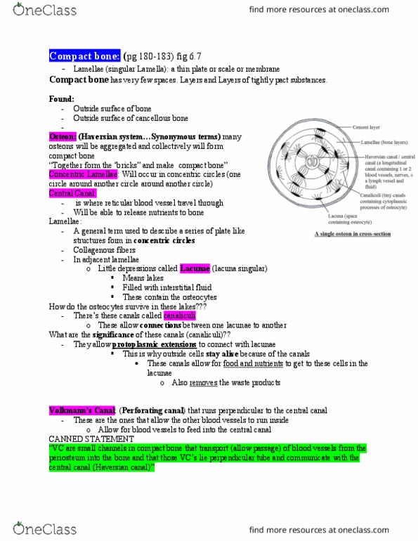
outside surfaces of other bones. Compact bone consists of cylindrical units called
osteons. Each osteon contains concentric lamellae (layers) of hard, calcified
matrix with osteocytes (bone cells) lodged in lacunae (spaces) between the
lamellae. Smaller canals, or canaliculi, radiate outward from a central canal,
which contains blood vessels and nerve fibers. Osteocytes within an osteon are
connected to each other and to the central canal by fine cellular extensions.
Through these cellular extensions, nutrients and waste are exchanged between
the osteocytes and the blood vessels. Perforating canals provide channels that
allow the blood vessels that run through the central canals to connect to the
blood vessels in the periosteum that surrounds the bone.
• Spongy bone consists of thin, irregularly shaped plates called trabeculae,
arranged in a latticework network. Trabeculae are similar to osteons in that both
have osteocytes in lacunae that lie between calcified lamellae. As in osteons,
canaliculi present in trabeculae provide connections between osteocytes.
However, since each trabecula is only a few cell layers thick, each osteocyte is
able to exchange nutrients with nearby blood vessels. Thus, no central canal is
necessary.
Document Summary
There are two kinds of bone tissue): compact bone is the hard material that makes up the shaft of long bones and the outside surfaces of other bones. Compact bone consists of cylindrical units called osteons. Each osteon contains concentric lamellae (layers) of hard, calcified matrix with osteocytes (bone cells) lodged in lacunae (spaces) between the lamellae. Smaller canals, or canaliculi, radiate outward from a central canal, which contains blood vessels and nerve fibers. Osteocytes within an osteon are connected to each other and to the central canal by fine cellular extensions. Through these cellular extensions, nutrients and waste are exchanged between the osteocytes and the blood vessels. Trabeculae are similar to osteons in that both have osteocytes in lacunae that lie between calcified lamellae. As in osteons, canaliculi present in trabeculae provide connections between osteocytes. However, since each trabecula is only a few cell layers thick, each osteocyte is able to exchange nutrients with nearby blood vessels.