01:830:101 Lecture Notes - Lecture 7: Visual Cortex, Occipital Lobe, David H. Hubel

21 Apr 2016
School
Department
Course
Professor


172
01:830:101 Full Course Notes
Verified Note
172 documents
Document Summary
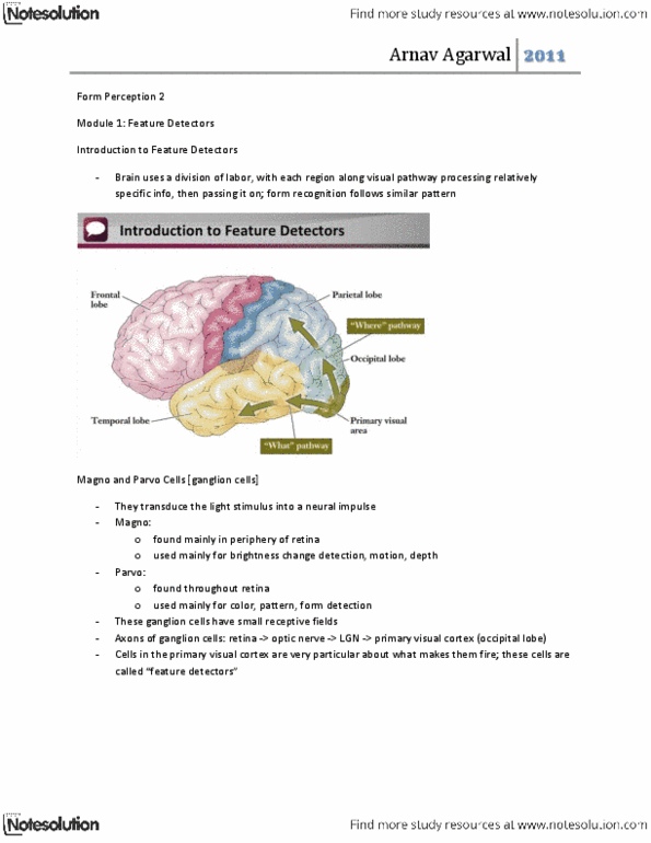
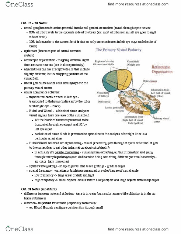
Information crosses over to other hemisphere of the brain. David hubel and wiesel discovered that we have a topographic map in the occipital lobe (primary visual cortex). Interested in just studying visual perception in cats. Use invasive single cell techniques, open cat skulls, attach recording device to. To see what kind of and where info is being processed-- receptors to recognize all. Color receptors are antagonistic- like red and green receptors. center responds to cells orientation of lines one form of light. Additive color mixing- adding wavelengths of light to a color, changes. (ex, all of them= white) Subtractive- removal of wavelengths of light, alters appearance of colors (ex, all of them=brown) 1800s, we have 3 diff receptors for color mixing located in the brain. Right: we have receptors for color vision (cones) Wrong: cones are in eye, not the brain. it processes in the brain. Red green, blue yellow, black white receptors. one in the center, one surrounds.