01:119:116 Lecture Notes - Lecture 2: Crystal Violet, Gram Staining, Cell Membrane

14 Mar 2016
School
Department
Course
Professor
Document Summary
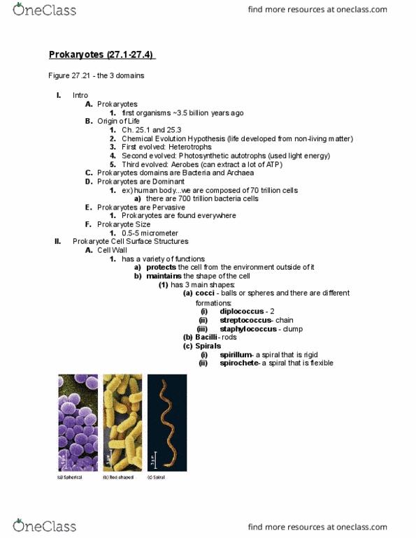
Intro: prokaryotes: first organisms, 0. 5-5 microns, chemical evolution hypothesis. Cell surface structures: functions, protects, cell shape. Cocci : single, group of 2 -> diplococcus, chain - streptococcus, clump - staphylococcus. Spirals: rigid = spirillum, flexible = spirochete: prevents bursting in hypotonic environment. Figure 7. 12 for all examples: peptidoglycan, polymer: sugars cross-linked, only domain bacteria, not in archea, eukaryotes - plants = cellulose, fungi = chitin. Gram stain: 2 stains, crystal violet -> purple color, safranin -> pink color. Gram positive: thick cell wall peptidoglycan, crystal violet retained by cell, therefore purple. Gram negative: thin peptidoglycan layer, does not retail crystal violet, therefore safranin not masked-> pink, outer membrane lipopolysaccharide (lps): carbs and lipids, toxic -> fever, differentiate -> correct antibiotics. 2: capsules and slime layers, polysaccharides and protein, surround cell wall, capsule: organized figure 27. 4, slime layer: loose attachment, protective - dehydration, phagocytosis, fimbriae and pili figure. 27. 6: hair-like, attachment, endospores figure 27. 5, dormant stage, very resistant.