01:119:115 Lecture Notes - Lecture 2: Crystal Violet, Chemotaxis, Dna Replication

8 Jul 2015
School
Department
Course
Professor
Document Summary
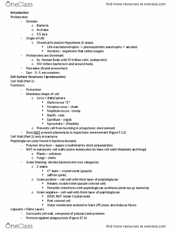
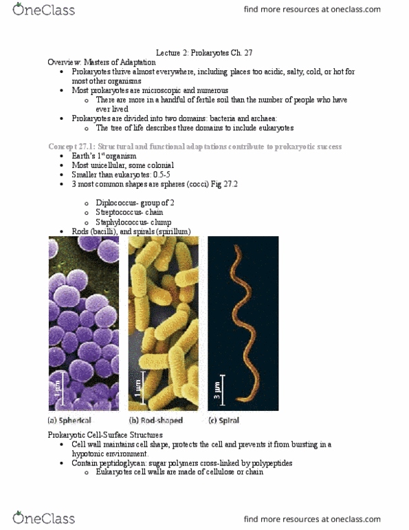
Three common shapes: spherical cocci bacteria , rod-shaped bacilli bacteria , spiral spirochetes , figure 27. 2. Prokaryotes have a cell wall: function of wall: In hypertonic environment cell shrivels (plasmolysis: structure of cell wall. Primarily peptidoglycans (mixture of sugars cross-linked by short polypeptides) Plants and fungi walls made from cellulose or chitin. Ii gram-staining: technique that allows differentiation of prokaryotes based on cell wall composition: stain with crystal violet dye + iodine, rinse chemicals off with alcohol, stain with red dye (safranin) Peptidoglycan traps crystal violet: gram-negative prokaryotes. Lipopolysaccharides can be toxic to host: figure 27. 3. Cell wall surrounded by sticky polysaccharides or proteins: facilitate attachment to substrate or other cells, well defined layer called capsule (figure 27. 4, not so well defined layer called slime layer. Some prokaryotes can survive harsh conditions by producing endospores: copy of dna enclosed by multilayer coat, coat allows cell to remain dormant for long time.