PSYCH 3313 Lecture Notes - Lecture 12: Far-Sightedness, Grandmother Cell, Dichromacy

3 Oct 2016
School
Department
Course
Professor
Document Summary
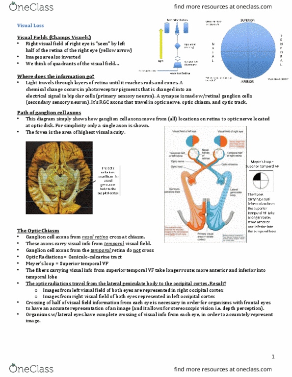
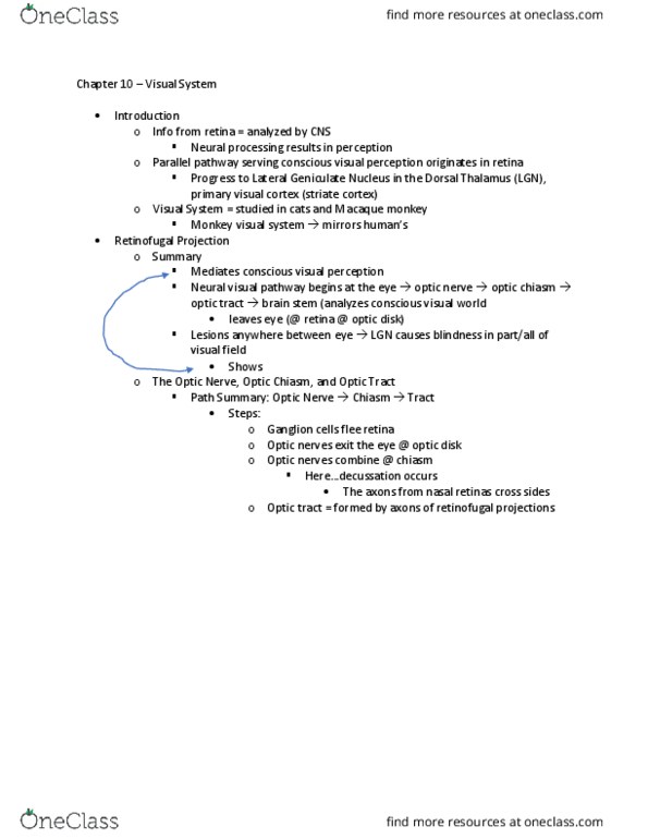
Visual fields: visual field (vf) = part of environment registered on retina. Right vf processed in left hemisphere (not just right eye) Left vf processed in right hemisphere (not just left eye: nasal view of each eye goes contralateral, temporal view of each eye stays ipsilateral. Ganglion cell axons bundle together and exit each eye through the optic disk, forming an optic nerve (cn ii) leaving each eye. About 50% of fibers cross to opposite hemisphere at optic chiasm. Small number of retinal axons: superior colliculus. 10% retinal axons: lateral geniculate nucleus (lgn) Projects to primary visual cortex (v1), visual perception. Pathways to the brain: right and left visual field to retina, optic nerve (ii) to optic chiasm to optic tract. [10%] superior colliculus and other secondary midbrain nuclei: [90%] lateral geniculate nucleus (lgn) of thalamus, optic radiations to primary visual cortex (v1) Pathways of visual analysis in the lgn of the thalamus: magnocellular.