MARKET 1 Lecture Notes - Lecture 3: Intercostal Arteries, Hiatus Hernia, Internal Intercostal Muscles

Document Summary
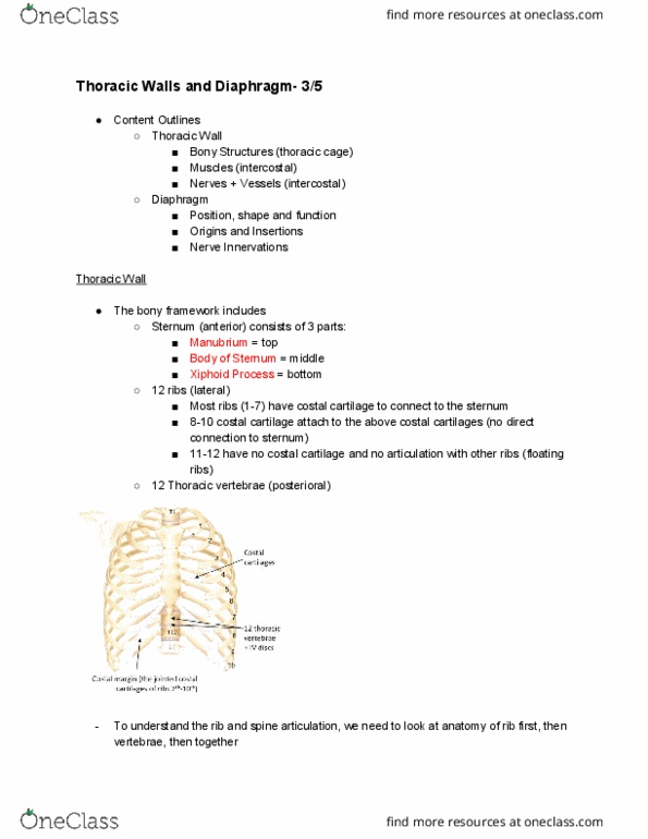
The thorax is the area located between the neck and the abdomen. The thoracic cavity contains the lungs and heart as well as the major blood vessels, part of the trachea and the oesophagus. These contents are protected by the thoracic wall. The thoracic wall consists of the sternum, the thoracic vertebrae, the 12 pairs of ribs and costal cartilages, the intercostal muscles and the arteries and nerves that supply the wall. The thoracic cage is composed of bone and cartilage and it provides flexibility and rigidity. The costal cartilages extend the ribs anteriorly and attach to the sternum. There are three types of ribs: true ribs- costal cartilage attaches directly to the sternum. False ribs- costal cartilage attaches to the cartilage above the sternum. Floating ribs- they do not attach to the sternum at all. The first rib that you can feel is the second rib.