BIOL 120 Lecture Notes - Lecture 21: Internal Urethral Orifice, Lamina Propria, Urinary Bladder

Document Summary
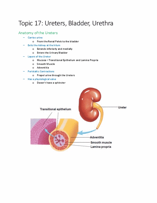
Vascular ct + lymphatic vessels + nerves. Transport urine from renal pelvis to urinary bladder via peristalsis. No anatomical valve @ opening of ureter into bladder. As urinary bladder fills w/ urine pressure compresses oblique openings into the ureters prevents backflow of urine. Small triangular area in floor of bladder. 3 layers of smooth muscle fibers: inner longitudinal. @ opening to urethra => circular fibers form internal urethral sphincter (involuntary) inferior => external urethral sphincter. @ posterior + inferior surfaces => areolar ct. Over the superior surface => serosa (layer of the. = discharge of urine from urinary bladder. Stretch receptors in bladder wall are triggered when volume exceeds 200- 400 ml stretch receptors transmit nerve impulses unto spinal cord micturition center micturition reflex triggered. => discharges urine from urinary bladder via parasympathetic impulses that cause contraction of the detrusor muscle + relaxation of the internal urethral sphincter muscle (voluntary)