BSC 1086C Lecture Notes - Lecture 1: Interatrial Septum, Papillary Muscle, Intercostal Space

Document Summary
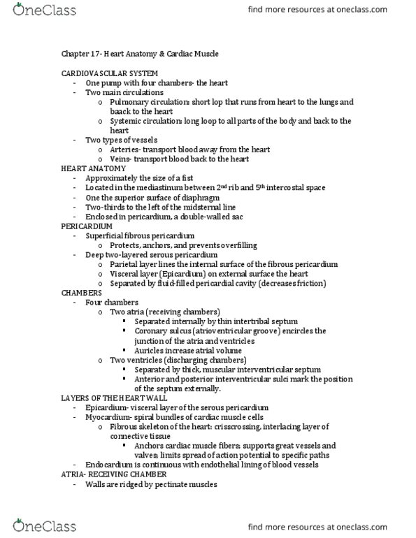
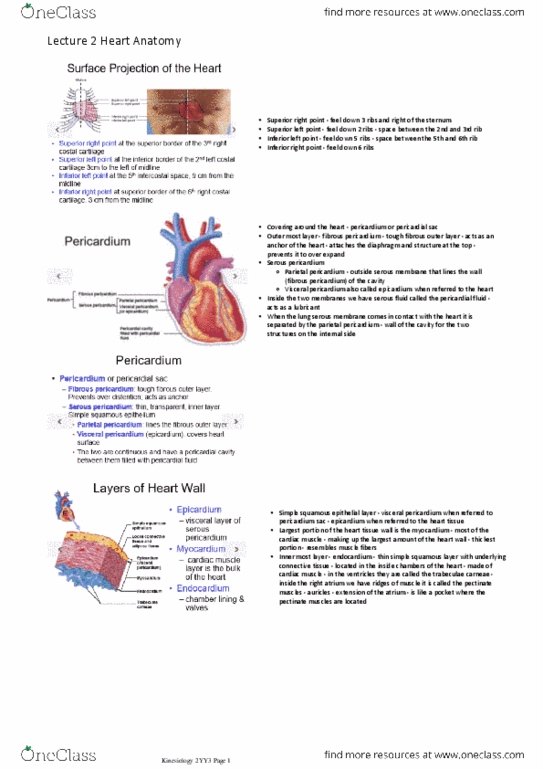
Chapter 17- heart anatomy in-class notes: the heart is approximately the size of a fist. It is located in the mediastinum between the second rib and fifth intercostal space. 2/3 of the heart is located to the left of the midsternal line. It has a tough outer layer called superficial pericardium. Its purpose is to anchor cardiac muscle fibers, to support vessels and valves and to limit the spread of action potentials: the heart has 4 chambers: two receiving chambers called atria and two discharging chambers called ventricles. The receiving chambers have appendages called auricles that increase the atrial volume if needed: the atria are separated internally by the interatrial septum and the ventricles by the interventricular septum. The atria are found above this sulcus and the ventricles below: the thin walls of the atria are ridged by the small pectinate muscles, whereas the walls of the ventricles show big muscle ridges called trabeculae.