BIOSC-101 Lecture Notes - Lecture 19: Color Vision, Choroid, Cornea

14 Sep 2020
School
Department
Course
Professor
Document Summary
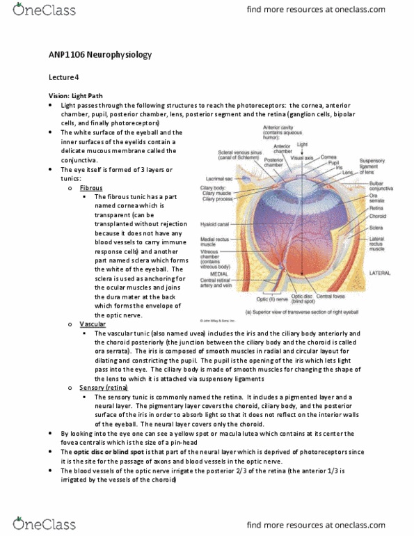
Consists of blood vessels, lymphatics, and intrinsic eye muscles. Regulates the amount of light entering the eye. Secretes and reabsorbs jelly-like fluid (aqueous humor) Function: contains blood vessels that supply eye tissue with nutrients & oxygen contains melanin, which absorbs light reflected from the retina. Function: ring of muscle tissue that encircles the lens holds lens in place and changes its shape. Function: allows light to enter the eye & reach the retina. Dilates in dim light; constricts in bright light. Location: opening in the center of the iris. Location: muscular part of the choroid in front of the ciliary body. Function: provides fine focusing of the light (accommodation) Made of two layers: (pigmented layer outer layer) / (neural layer inner layer) Retina cells: rods (night vision) and cones (color vision) Function: contains photoreceptors that respond to light by generating electrical signals. Region of the retina with greatest concentration of cones. Function: objects are focused here for sharp vision.