BIOL 211 Lecture Notes - Lecture 2: Syphilis, Scanning Electron Microscope, Protozoa

25 Aug 2016
School
Department
Course
Professor
Document Summary
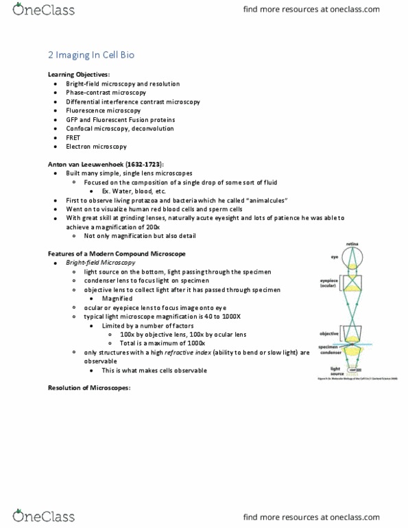
Anton van leeuwenhoek (1632-1723) -janitor/clothes merchant in delft, holland. Made no speculation about the significance: types of microscopy. 1000x magnification, stain for contrast (bacteria are mostly water: darkfield microscopy. Use: to visualize cells that might be distorted by staining or to visualize living cells. Treponema pallidum: bacterium that is the causative agent of syphilis. Only scattered light by object enters the objective lens: phase microscopy. Light waves going through and outside of object out of phase- leads to increased contrast. Use: observe structural detail (e. g. storage granules: fluorescence (uv) microscopy. Upon exposure of the dye to uv, there is readmission of visible light of a certain wave length. The object appears colored against a dark background. Use: diagnosis of infections caused by bacteria, viruses, protozoans. Laser beam scans various depths of a specimen to deliver a sharp image in a single plane. Use: can obtain a highly focused view at any level.