KINE 2031 Lecture Notes - Lecture 5: Supraspinous Ligament, Lambdoid Suture, Anterior Longitudinal Ligament

17 Oct 2016
School
Department
Course
Professor

Document Summary
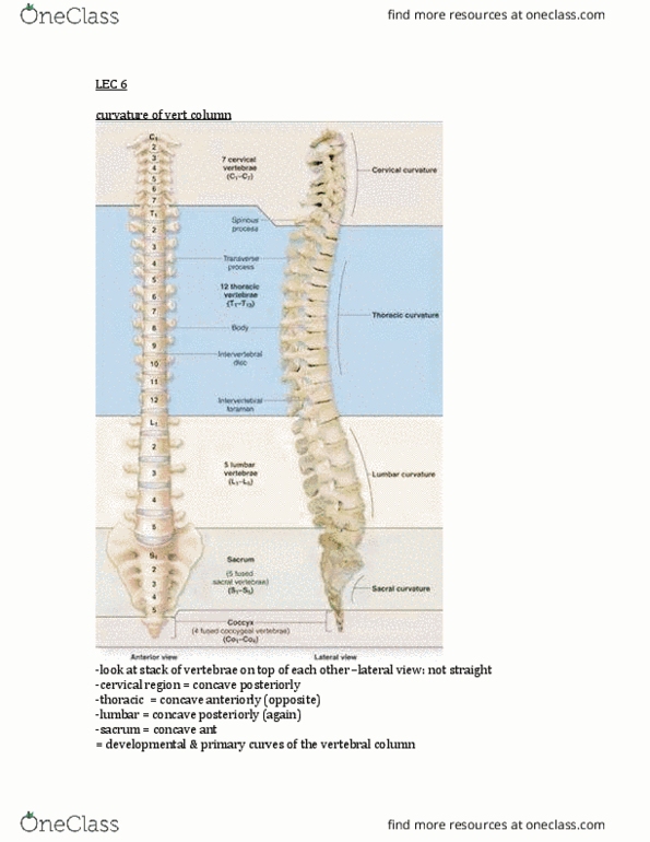
*squamous = flat cells, thin flat part of temporal bone at that region. Vertebral column: series of bones stacked on top of eachother. Thoracic next 12 down (designed for attachment of ribs) Sacrum 5 fused together in triangular shape. Diff shape & sizes but same parts, beside the 1st cervical vertebrae*: Hole = vertebral foramen to allow passage of sc to travel down. Pedicle = joins body to transverse process on either size (little feet) Lamina = joining to transverse process to spinous process. Superior surface = for stacking bone on eachother. Inferior surface (articulates with bone above) = stacking bones. When u stack 2 vertebrae on top of eachother, you get the intervertebral foramen: opening --hole to allow spinal nerves to exit on either side of the spinal cord. Has an interior arch & bigger posterior arch. Hole = transverse foramen: vertebral artery travel up & in cranial cavity to supply blood to brain **only in cervical bones!