Physiology 3140A Lecture Notes - Lecture 10: Calcium Atpase, Rod Cell, Chromophore

22 May 2018
School
Department
Course
Professor

Physiology 3140
Dr. Rylett
Lecture 10
Regulation of cGMP Phosphodiesterase: Role of cGMP in Signal Transduction in the Visual System
Anatomy of Retinal Rod Cell
- There are 4 basic parts of the retinal rod cell:
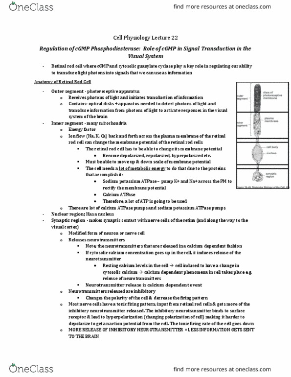
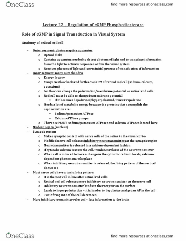
- outer segment:
o contains all the photoreceptive apparatus
o has the optical discs in it
o contains the apparatus needed to detect photons of light as well as transduce info from
those photons to be able to actually activate responses within the visual system in brain
- inner segment:
o many mitochondria
o its like an energy factory
o there is going to be a lot of ion flow back and forth across the PM of the retinal rod cell,
this can change the polarization (aka the membrane potential) of each retinal rod cell
o the retinal rod cell has to be able to change its membrane potential and to do this, it
needs a lot of metabolic energy bc some of the proteins thats going to be able to help do
this is Na+/K+ ATPase and Ca2+ ATPase
o there are a lot of the Ca2+ ATPase and Na+/K+ ATPase pumps in the inner segment
- nuclear region
o has the nucleus
- synaptic region:
o makes synaptic contact with nerve cells of the retina and along the way to the visual
cortex
o this is a modified form of a neuron
o releases neurotransmitters from here
o these neurotransmitters are released in a Ca2+ dependent fashion
▪ if the cytosolic Ca2+ concentration increases, more
neurotransmitter will be released
o these neurotransmitters are INHIBITORY – this will change the
polarity of the next cell and decrease the firing pattern of the next
cell
▪ most nerve cells have a tonic firing pattern – if it gets
inhibitory neurotransmitters from the retinal rod cell, then
the inhibitory neurotransmitters will bind to the receptors
on the surface, leading to hyperpolarization (change the
polarization of the cell)
▪ this makes it harder to depolarize and get an AP
▪ Then the tonic firing rate of the nerve cell will decrease
find more resources at oneclass.com
find more resources at oneclass.com

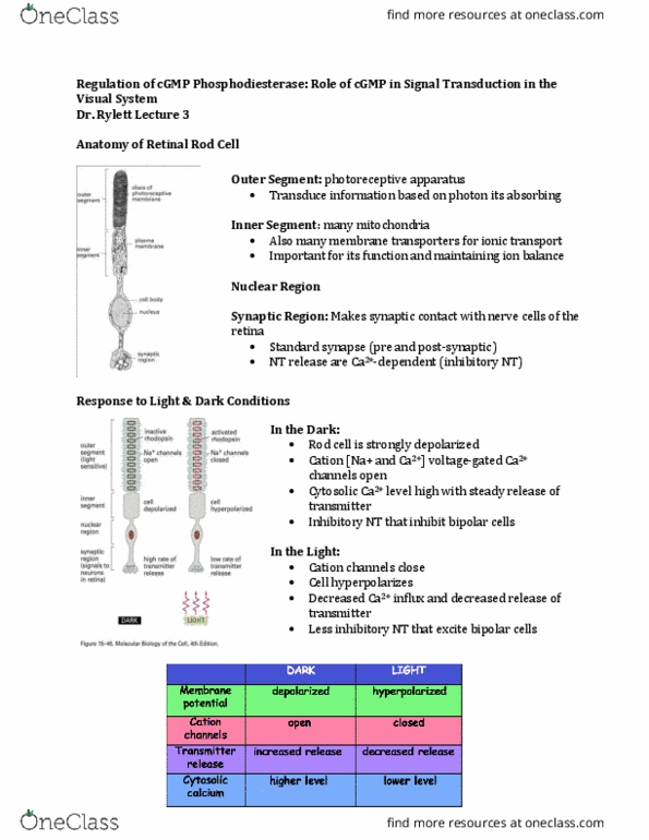
Responses to Light and dark conditions
- Note: in the image it says Na+ channels, but its actually a cation channel (they are selective to
allow both Na+ and Ca2+ through)
- rhodopsin (a GPCR) is the receptor that is going to detect photons of light
- rhodopsin has a portion of the molecule which is a retinal chromophore
o so a portion of the receptor, which is same as the ligand binding site, is an adapted form
of vitamin A
- the retinal chromophore can detect photons of light
o when photons of light hit the rod cell, they change the conformation/energy state of the
retinal, this is going to couple a G protein, which is going to couple an effector
- in the dark:
o rhodopsin is INACTIVE
o rod cell is strongly depolarized
o cation [Na+ and Ca2+] voltage-gated Ca2+ channels open
o cytosolic Ca2+ level high with steady release of transmitter
o no light - cation channels are open and gated – Cations flow down electrochemical
gradient (outside is positive inside is negative)
o so the Na+ causes the retinal rod cell to be depolarized relative to the RMP
- light causes:
o photons of light hit rhodopsin and activate it through conformational change of the
retinal chromophore
o the Na+ and Ca2+ channels spontaneously start to close,
o cell hyperpolarizes
▪ recall: there is lots of Na+/K+ ATPase in the inner segment trying to get
membrane potential back to RMP
▪ and bc the cation channels are closed, the Ca2+ that is in there is getting
pumped out by Ca2+ ATPase
o decreased Ca2+ influx and decreased release of transmitter
find more resources at oneclass.com
find more resources at oneclass.com

Transmitter inhibits postsynaptic neurons
- illumination(light) frees the next nerve cell that the retinal rod cell synapses on from inhibition
and thus excites them
- causes it to increase to its tonic firing rate = excitation
- the rate of transmitter release from rod cells is graded to light intensity
o little bit of light = few rhodopsin molecules affected = small # of neurotransmitter
released
o lot of light = more rhodopsin molecules affected = large # of neurotransmitter released
o humans are able to detect light over a broad range
o for this to be able to happen, our visual system at the level of the retinal rod cell has to
be able to respond over a very broad range
o its a graded intensity !!!!
Rod cells contain visual pigment rhodopsin
Rhodopsin has a light-absorbing portion of complex retinal (vitamin A):
- retinal chromophore(form of vitamin A) is the portion of rhodopsin that detects like
- absorption of light causes conformational change in retinal and cascade of events involving
cGMP
o conformational change is coupled to a G protein which is coupled to an effector
o the effector is cGMP phosphodiesterase
- this is only light-dependent step in vision
o ex: if you have a defect where you are deficient in vitamin A or retinal, so your visual
system doesnt have that chromophore associated with rhodopsin
o you would not be able to detect differences in light
o therefore it is important that this is the ONLY light dependent step
cGMP is important regulatory molecule in photosensitive rod cell
- light on rod cells changes intracellular Ca2+, leading to activation of G protein , this leads to Ca2+
changes and decrease cGMP
- large decrease in cGMP - critical event in transduction of response to light
Cellular mechanisms regulating light perception
- in darkness:
o level of cGMP in rod cells is high because activity of cGMP-phosphodiesterase is low
o no photons of light hitting the retinal chromophore- not coupling G protein – not
coupling the effector = phosphodiesterase activity is low →this leaves any cGMP in the
cell where it is
- activation of retinal pigment molecules by light:
find more resources at oneclass.com
find more resources at oneclass.com
Document Summary
Regulation of cgmp phosphodiesterase: role of cgmp in signal transduction in the visual system. In the dark, in retinal rod cells, cgmp is high and bound to cation channels, ca2+ and. Pm and the outside of the plasma membrane is facing the patch pipette. We can test to see what kind of solutions change the conductance of these cation channels. So,this is a good experiment to determine what the ligand is that causes the opening: this is an exam question! Inside-out patch of membrane from outer segment of rod cell for measurement of changes in current flow under voltage-clamp conditions. Expose patch of membrane to solutions that contain cgmp. With cgmp, get an increase in current flow this is the (cid:498)dark current(cid:499) Embedded in that, we have the gpcr, rhodopsin, which has the visual pigment, retinal (which is. In the outer segment of the retinal rod cell, we have a disc like membranous structure able to detect photons of light)