Physiology 3140A Lecture Notes - Lecture 4: Focal Adhesion, Paxillin, Stress Fiber

2 May 2018
School
Department
Course
Professor

Physiology 3140 Lecture 4
Cell-Matrix Interactions 2
September 15, 2017
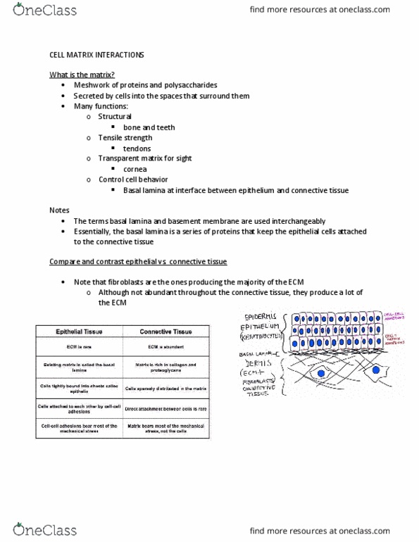
- Dermis is structure in skin that has connective tissue: ECM + fibroblasts + collagen + multi-adhesive
proteins
o Connective tissue around bone is different!
- Absorptive or secretory epithelium = one layer thick
- Squamous epithelium = many layers
Resolution power of Immunofluorescence microscopy
- Approximately ½ the wavelength of light
- For e.g., assuming visible light is 400-600 nm, resolution is 200-300 nm
o Use immunofluorescence because have many different wave lengths
- Therefore, two objects closer than 200-300 nm will superimpose (i.e., they will look like one object)
o Remember! RESOLUTION = ½ WAVELENGTH
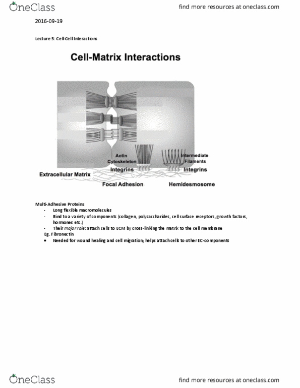
Cell-Matrix Interactions
- Focal adhesions are transient
Focal Adhesions in quiescent or migratory lung tumor cells
find more resources at oneclass.com
find more resources at oneclass.com

- Migratory lung tumor cells
o Started with lung tumor cells, monolayer growing on cover slip, fixed, permeabilized,
primary antibody (Paxillin)
- Paxillin is a protein that labels focal adhesions
- Actin cytoskeleton
o Focal adhesions: integrin bound to actin
- Green channel: antibody paxillin
- Red channel: compound that fluoresces red that binds to actin cytoskeleton
- Cortical staining goes around the cell
- Stimulated now they are migratory
o Takes 24 hours to see
o Paxillin stain: the size of the focal adhesions are much larger
▪ Cells need traction in order to move – make bigger focal adhesions (suction cup) so
they can grab onto the plate (culture place or cover slip)
o Normally, in a cell the actin is all around the cell CORTICAL
o When you migrate, your actin becomes your arms – what you grab on to the plate through
the focal adhesion and go along
▪ STRESS FIBERS
▪ Actin rearranges depends on what’s happening inside of the cell
- Look at the overlay:
o Dragging itself along the plate
o Can still see red and green in the merge because stress fibers and focal adhesions do not co-
localize perfectly!
▪ They are not closer than 200nm
▪ Where it is yellow: they are closer than 200nm this is the attachment point
between the stress fiber and the focal adhesion
Cell junctions
- There are 3 types of junctions in the cell
o Anchoring junctions
• Cell-cell
• Cell-matrix
• Mechanically attach cells to their external surroundings
o Occluding junctions
• Cell –cell
o Communication junctions
• Cell – cell
- Involved in transmitting information back to the cell
o One cell produces ECM because of what is happening to it, the cell next to it will see changes
in the ECM and will responds to it
• Responds to it by having a relay of proteins and structures going from the ECM all
the way to the nucleus
find more resources at oneclass.com
find more resources at oneclass.com

Transmission of Information
- Cell is upside down
o ECM is on top in the image
- Plasma membrane transmembrane proteins
o E.g. integrin
o Physical interact and bind ECM
o Associated with other proteins (e.g. plectin, vinculin)
• Adaptor proteins at the cell surface
- As you go into the cell, toward the nucleus
o Cytoskeleton = actin
o Actin cytoskeleton will bind to its adaptor proteins on the nuclear membrane
- Nuclear structural proteins
o E.g. lamin
o Inside of nucleus that feel or respond to rigidity of the cytoskeleton and respond to it
o When proteins are mutated, get: laminopathies
Human connective tissue diseases
- Arthritis (osteoarthritis)
o Affects articular cartilage, joint, underlying bone
- Scleroderma
o Autoimmune disease that attacks parts of the skin
o Causes scaring
o Epithelium gets thin, get unwanted tissue repair within the dermis
o Skin condition
- Vitamin C deficiency (Scurvy)
o Bleeding gums
• Connective tissue in your gums break down because your collagen breaks down
• Teeth fall out because they can not hang on to the gums
o Vitamin C is VERY important for good collagen production
o Ends up everywhere else, wherever you have collagen will be affected
- Osteogenesis Imperfecta
o Bone disease
find more resources at oneclass.com
find more resources at oneclass.com
Document Summary
Dermis is structure in skin that has connective tissue: ecm + fibroblasts + collagen + multi-adhesive proteins: connective tissue around bone is different! Absorptive or secretory epithelium = one layer thick. For e. g. , assuming visible light is 400-600 nm, resolution is 200-300 nm: use immunofluorescence because have many different wave lengths. Therefore, two objects closer than 200-300 nm will superimpose (i. e. , they will look like one object: remember! Focal adhesions in quiescent or migratory lung tumor cells. Migratory lung tumor cells: started with lung tumor cells, monolayer growing on cover slip, fixed, permeabilized, primary antibody (paxillin) Paxillin is a protein that labels focal adhesions. Actin cytoskeleton: focal adhesions: integrin bound to actin. Red channel: compound that fluoresces red that binds to actin cytoskeleton. Look at the overlay: localize perfectly: they are not closer than 200nm, where it is yellow: they are closer than 200nm this is the attachment point between the stress fiber and the focal adhesion.