Physiology 3120 Lecture Notes - Lecture 43: Endochondral Ossification, Type Ii Collagen, Intramembranous Ossification

15 May 2018
School
Department
Course
Professor

Lecture 43 – Bone Physiology
Hormonal regulation of calcium balance
- Bones are regenerative, constantly modified and not static
- Entire skeletal regenerates itself within 6-7 years (skeleton remodels into a new skeleton)
- Calcium deposits into our bone and is stored there for later - strengthens our bones
- Parathyroid hormone regulates the plasma level of calcium in the extracellular fluid volume
by conserving calcium from the distal convoluted tubule
o Reabsorb more calcium because of PTH = elevating blood calcium levels
- Maintain calcium homeostasis by reabsorption and absorption
- Calcitrol (vitamin d) affects absorption of calcium in the gastrointestinal tract
o Can enhance calcium absorption
- 99% of body calcium is stored in bones
o Calcium forms a mineral that strengthens bone tissue
o It is an easy access point
o If ever need to increase plasma calcium levels, PTH stimulates bone to degrade to
release calcium into blood
o Can degrade bone to release calcium when necessary don’t want it too quick
- Calcium is stored as hydroxyapatite crystals (calcium and phosphate salts) in bone to
prevent large changes in plasma calciums levels
o Inorganic material is part of the collagen matrix that is formed
o By storing calcium in bone, it prevents large increases in plasma calcium
o Crystals provide bone strength (inorganic component of bone)
- Bone is composed of:
o Type I collagen
o Hydroxyapitate crystals
- Bone serves more functions than just locomotion and protection of vital organs
o Bone is not just for locomotion
o Skeletal system serves as an endocrine organ
o Controls ion balance
o Maintains calcium to ensure everything functions properly
Types of bone formation
- Two types of bone formation in embryo when developing skeleton of body:
o Endochondral ossification **most bones form this way (femur, tibia, long bones)
▪ In utero, cartilage template was replaced by bone
▪ Bones continue to grow because of remaining cartilage template
▪ Growth plate: cartilage template the remains so bones can be longer
o Intramembranous ossification
▪ Mesenchymal cells in utero become bone directly
▪ Cells become bone forming cells (osteoblasts – lay down matrix and
inorganic matrix) → no cartilage template
find more resources at oneclass.com
find more resources at oneclass.com

- Bones are categorized by the type of developmental process that forms them
- Bones are continuously remodeled
Composition and organization of bone
- Bones of the skeleton have different processes that they underwent to be developed
- Long bones (femus), tibia, ulna formed by endochondral ossification
- Bones of skull and sternum formed my intramembranous ossification
- Trabeculae/spongy bone has gaps where bone marrow is
- Chondrocytes (cells of cartilage) maintain a healthy extracellular matrix rich in
proteoglycans and type II collagen
o There are not many cells are in articular cartilage but chondrocytes are responsible
for maintaining a healthy ECM
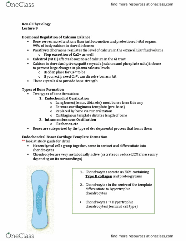
Endochondral bone: cartilage template formation
- 1. Chondrocytes secrete an extracellular matrix containing type II collagen and
proteoglycans
o In utero in early development, cartilage template is formed first
o Chondrocytes come from mesenchymal stem cells
o ECM secreted is almost identical to articular cartilage
o Type II collagen is scattered in its formation
- 2. Chondrocytes in the centre of the template differentiate to hypertrophic
chondrocytes
o Hypertrophic chondrocytes: larger chondrocytes that increase in size and they are
differentiated (maturing from a chondrocyte)
o They secrete a different matrix
find more resources at oneclass.com
find more resources at oneclass.com

- 3. Hypertrophic chondrocytes secrete type x collagen and less proteoglycans
o Type X collagen: found in hypertrophic region (diff. type of collagen is secreted)
o Collagen provides strength while proteglycans provide cushion
o More collagen = more strength to the matrix
o Less proteoglycans = less squishability/compression of the matrix
o Strength is increased where the hypertrophic chondrocytes are (center of the
template)
- 4. Localized hypoxia in hypertrophic zone of the template triggers secretion of
angiogenic factors (VEGF)
o Hypertrophic chondrocytes are metabolically active cells
o Chondrocytes receive nutrition from diffusion (oxygen, glucose, etc.)
o Hypoxia develops because the template gets too big
o There is localized decrease in oxygen in the template can’t get far enough into the
template)
o Hypertrophic cells secrete VEGF– stimulates the recruitment of blood vessels
(angiogenic factor)
o If hypoxic and cells require more nutrition/oxygen, want to deliver blood cells to
region to provide proper nutrition for the metabolically active cells
find more resources at oneclass.com
find more resources at oneclass.com
Document Summary
Bones are regenerative, constantly modified and not static. Entire skeletal regenerates itself within 6-7 years (skeleton remodels into a new skeleton) Calcium deposits into our bone and is stored there for later - strengthens our bones. Parathyroid hormone regulates the plasma level of calcium in the extracellular fluid volume by conserving calcium from the distal convoluted tubule: reabsorb more calcium because of pth = elevating blood calcium levels. Maintain calcium homeostasis by reabsorption and absorption. Calcitrol (vitamin d) affects absorption of calcium in the gastrointestinal tract: can enhance calcium absorption. 99% of body calcium is stored in bones: calcium forms a mineral that strengthens bone tissue. If ever need to increase plasma calcium levels, pth stimulates bone to degrade to release calcium into blood: can degrade bone to release calcium when necessary (cid:523)don"t want it too quick(cid:524) Calcium is stored as hydroxyapatite crystals (calcium and phosphate salts) in bone to prevent large changes in plasma calciums levels.