Medical Sciences 3999A/B/Y Lecture Notes - Lecture 25: Intestinal Villus, Gastrointestinal Tract, Muscularis Mucosae

20 Feb 2020
School
Department
Professor
Document Summary
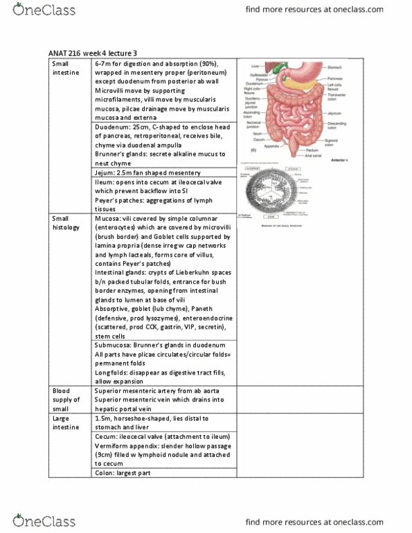
Lecture 25 - gi (small & large intestines) Main functions - to continue gastric digestion into the duodenum, to combine intestinal enzymes with pancreatic enzymes and bile to emulsify macromolecules, and to absorb digested food. Wall consists of four layers: mucosa, submucosa, muscularis and serosa. Histologic differences seen in mucosal and submucosal portions. Intestinal wall"s absorptive surface area amplified by tissue specialization of submucosa and mucosa - four degrees of folding: Permanent circular folds of mucosa and submucosa encircling the lumen. Appears ~5 cm distal to pylorus --> distinct where duodenum joins jejunum. Diminish in size progressively --> disappears halfway along ileum. Finger-like projections of mucosa covering entire surface of small intestine. Extends deep into mucosa --> forms crypts between adjacent villi. Simple tubular intestinal glands; also increases surface area. Formed by invaginations of mucosa between adjacent intestinal villi. Each enterocyte has several thousand packed microvilli (striated border) Lamina propria: surrounds intestinal glands and forms core of villi.