Kinesiology 2222A/B Lecture Notes - Lecture 13: Orbicularis Oculi Muscle, Occipitalis Muscle, Facial Nerve

17 Oct 2018
School
Department
Course
Professor

- muscles innervated by same nerve share the same function because derived from same
embryonic origin (share nervous supply)
- every muscle has a primary function (thats what we need to know)
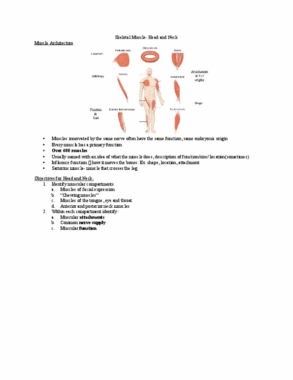
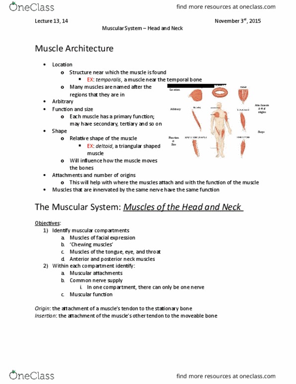
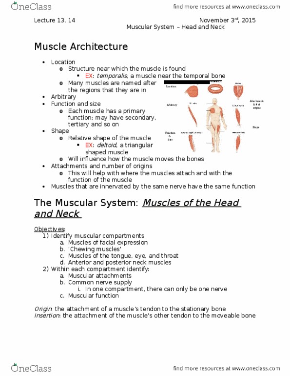
Muscle Architecture:
- over 600 muscles
- classify based on function and size
- sometimes classify based on location
- some are classified based on attachment and origins
- shape influences function of the muscle
- some isolated cases of classifying
- muscles of the face:
- muscles of facial expression
- muscles of chewing
The Muscular System:
- think function of muscles
- large muscles often have multiple functions
- not always using all the fibres of the muscle to full force of contraction
- can decide different movements based on which muscles you want to contract
Facial Expression: innervated by facial nerve
1. a) muscles of facial expression
- muscles attach to bone, skin, and fascia (supporting skin) so you can move the skin for
expressions of the face
- origins are the base for the muscle: pulling point. in red
- insertion is whats being pulled (towards origin) in blue
- majority to soft tissues of skin
- Epicranius: fascia. if you raise eye brows, can feel it moving. similar to tendon. imbedded in
centre of large muscle with 2 bellies on opposite ends of fascia:
- frontalis belly: will raise eyebrows if selectively contracted
- occipital belly: posterior
- orbicularis oculi: cirrcular, goes around each eye, boney
- contraction causes the eye to close, known as the blinking muscle
- Zygomaticus Major and Minor: originates from zygomatic process to corner of mouth
- major is larger than the minor
- run parallel to each other at same location at corner of the mouth (can see gap)
- known as the smiling muscles (zygomatic process to corner of mouth) bring corners of
mouth up forming a smile
- Orbicularis Oris: around the mouth. “kissing muscle”
- pursing lips together

- Risorius: mainly attached to soft tissues and skin
- grimacing muscle: corners of mouth outwards
- Buccinator: whistling muscle
- draws teeth tight against cheeks
- grouped with facial expression
- involved in muscles of chewing as well
- helps move the jaw from side to side (trying to keep food in b/w molars)
- innervation is the same as the rest of the facial expression muscles
- Depressor Anguli Oris: attaches to corner of the mouth
- on a downward angle knows as frowning muscle
- Platysma: covers the anterior neck
- very superficial
- called the shaving muscle
- tenses the skin on the anterior neck
Facial Nerve:
- All these muscles innervated by cranial nerve #7- facial nerve
- arrises on brain stem
- enters from cranial cavity into internal acoustic metus (IAM)
- on temporal bone
- leads into auditory canal on temporal bone
- one exits out of stylomastoid foramen b/w mastoid and styloid process of temporal
- once exits the skull, exposed to and runs right through parroted gland:
- salivary gland for eating
- splits into 5 brances: extending to different regions of the face
1. Temporal
2. Zygomatic
3. Buccal
4. Mandibular
5. Cervical
- each branch responsible for certain muscles of facial expression
- if any infection of parroted gland (duct enters oral cavity so it can happen) it will swell
making it risky for the facial nerve. swelling puts pressure on the facial nerve and can
hinder its activity
- Bell’s Palsy: partial or temporary damage to the facial nerve as result of infection to
parodied gland. wake up one morning with half the face feeling melted and unable to
innervate muscles. could be only one gland effected and could be only 1 side of the face.
80% is curable/treatable. some damage can be permanent to the nerve
Muscles of Mastication: active chewing
- many much larger and directly attach to bone (solid attachment to mandible)
Document Summary
Muscles innervated by same nerve share the same function because derived from same embryonic origin (share nervous supply) Every muscle has a primary function (thats what we need to know) Some are classified based on attachment and origins. Not always using all the fibres of the muscle to full force of contraction. Can decide different movements based on which muscles you want to contract. Facial expression: innervated by facial nerve: a) muscles of facial expression. Muscles attach to bone, skin, and fascia (supporting skin) so you can move the skin for expressions of the face. Origins are the base for the muscle: pulling point. in red. Insertion is whats being pulled (towards origin) in blue. Epicranius: fascia. if you raise eye brows, can feel it moving. similar to tendon. imbedded in centre of large muscle with 2 bellies on opposite ends of fascia: Frontalis belly: will raise eyebrows if selectively contracted. Orbicularis oculi: cirrcular, goes around each eye, boney.