Kinesiology 2222A/B Lecture Notes - Lecture 24: Subclavian Artery, Subclavian Vein, Brachial Plexus

16 Dec 2016
School
Department
Course
Professor

Anatomy Lecture December 1st
• A vessel will change names as it goes to different regions
• Systemic system
o Made up of veins and arteries
• Arteries
o Elastic walls
o High pressure
o Blood flow away from the heart
• Veins
o Thin walls
o Valves, could lead to pooling
o Low pressure
o Blood flow toward the heart
• Capillaries
o Do’t orr aout for this ourse
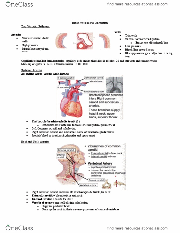
• Systemic arteries
o Starting with the heart
▪ Aorta
• Brachiocephalic trunk (brachium, and cephalic refers to the head)
o Branches into right common (further branches to come)
carotid, and the right subclavian space.
o Also have the left side
o These branches supply head and neck, upper limbs and
superior thorax
• Why we have a braccehpalic trunk
o This is like an extension cord, that is extending blood to
the right side which is how you get the right commono
carotid and subclavian
o Head and neck arteries
▪ The common carotid split furthers into the external and internal carotid
▪ External means external to the skull, and internal will go into the skull,
providing blood to the brain and other major centers.
o Vertebral artery
▪ Comes off the subclavian artery
▪ Small artery running in between the vertebral foramen
o Internal carotid and vertebral artery are responsible for giving blood to the brain
• Circle of Willis
o Vertebral artery from right subclavian and left subclavian, join to the circle of
willis. So will the internal carotids, the left and right.
find more resources at oneclass.com
find more resources at oneclass.com

o Thesee provide blood to the circle of willis.
o Anastomosis is common blood flow, when blood vessels form together and make
sure the brain is receiving blood
▪ Provides collateral blood flow
• Subclavian
o Subclavian artery moving into the subclavian space
o Intertwined with the brachial plexus
o When its moves distally turns into the axillary artery
o Left side is directly off the aortic arch
o Should be able to know the name of an artery based on a region
o Axillary provides blood to the pec, deltoid, scapular etc.
o As soon as it touches the humerous it is called the brachial artery
o Once past the elbow joint
• Splits into the ulnar and radial artery
o These go down the ulnar or radial side
o Astomoses occurs down in the hand, when these two arteries join together.
• As the arota continues
o Past the brachial saphalic trunk, its gives branches to the left common carotid
and left subclavian
o Also has branches going behind thew heart
o Giving branches to the abdominal/descending aorta
o Within the thoracic aorta, as the aorta descends, many branches give off giving
blood to the thoracic region
o All vessels are named according to which organ they provide blood to.
o E.g. esophageal artery provides blood to the esophagus
• Once in abdominal region
o More branches for reproductive and digestive organs
• Lets follow the descending aorta to the pelvis first
o Ends at around L4, the descending aorta splits into the left and right common
iliac
o This blood will now move towards the lower limbs.
o Common iliac
▪ Means more branches to come
• 3 unpaired abdominal arteries (everything so far has been paired with left and right)
• unpaired in the abdominal area
o 1. Celiac trunk
▪ has many branches
▪ many organs a part of digestion receive blood via the celiac trunk
▪ pancreas receive blood via the celiac artery
o Superior mesenteric
▪ Provides blood to most of the intestine
▪ Large and small
o Inferior mesenteric
find more resources at oneclass.com
find more resources at oneclass.com
Document Summary
Internal carotid and vertebral artery are responsible for giving blood to the brain: circle of willis, vertebral artery from right subclavian and left subclavian, join to the circle of willis. Celiac trunk: has many branches, many organs a part of digestion receive blood via the celiac trunk, pancreas receive blood via the celiac artery, superior mesenteric, provides blood to most of the intestine, large and small. On the medial side, brings blood back to the femoral vein: challenges, muscles around, cooling occurs here, too much cooling, valves can collapse, leading to exaggerated superficial veins, known as varacos veins. These veins rely on valves, especially ones down low. Superior vena cava: blood has unique way of bring blood back. Injections, intravenously, moves the drug into the systemic circuit, so everything you put in has access to all organ, blood will be filtered a(cid:374)(cid:455)(cid:449)a(cid:455)"s by kidneys and liver.