Kinesiology 1080A/B Lecture Notes - Lecture 3: Nerve Conduction Velocity, Alpha Motor Neuron, Luigi Galvani

11 Jun 2018
School
Department
Course
Professor

Nervous System
Divisions of Nervous System
- Two branches - CNS and PNS
- CNS- the brain and the spinal cord
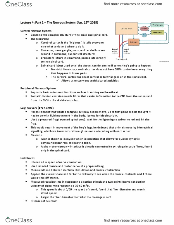
The CNS- the hierarchy
- Cerebral Cortex or Cerebrum is the big boss. Tells what to do and when to do
- Subcortical structures- thalamus, basal ganglia, pons and cerebellum
- Brainstem is third command
- Spinal cord is the slave system to all of the above
- There is no strict hierarchy, cerebral cortex does not have 100% of control
- Cerebral cortex also has direct control over spinal cord and might not need the subcortical
structures in the middle so the hierarchy isn't strict
Luigi Galvani- Animal electricity
- Before people thought people moved with movement of fluid
- Used a prepared frog
- Hooked a copper rod to lighting rod, lighting would struck and cause the frog’s legs to move
- Introduced the idea of Bioelectrical signalling
Neurons
- Causes Bioelectrical signalling
- Axon in the neuron has myelin sheath(insulation allows for speedy communication)
- People with MS have scars under myelin sheath
- Alpha motor neuron- interface directly with extrafusal muscle fibre (only in spinal cord )
Speed of nerve conduction
- Helmholtz (1850’s)
- Used isolated muscle and motor nerve of a frog
- Time between current and twitching of the leg
- At one point- current would be very close to cell body and Another stimulus the electrical current
would be farther down the axon
- Findings- Nerve conduction velocity is very fast, can be anywhere between 35-60 m/s (very fast).
Very important- eg. when falling we can recover very fast because of thats speed.
- Now humans can have speed upto- 100 m/s
- Depends on two things- how myelinated the axon is and diameter of the neuron
Diseases of the nerve
● ALS (amyotrophic Lateral Sclerosis) - influences amplitude of nerve conduction- alpha motor
neurons are damaged
● MS- influences the conduction speed, destroy and scar the myelin sheath, multiple scars along the
axon, impact neurons in brain, cortical structures and spinal cord
Many different types of Neurons:
find more resources at oneclass.com
find more resources at oneclass.com

● Motor (efferent) neuron
- transmit motor commands down the spinal cord
● Sensory (afferent) neuron
- transmits signals to, and up the spinal cord (to brain)
- These two areas are segregated in found in two different areas
How does info get from spinal cord to muscle?
Descending column leaves the spinal cord via the ventral root ganglion à extrafusal muscle fibers
How does info enter spinal cord from detection (spindles)?
Ascending column runs up the spinal cord à via the dorsal root ganglion
Cortical Structures:
● Cerebral cortex/cerebrum
o left hemisphere and right even
o parietal, frontal, occipital, temporal lobes
Phrenology:
● fMRI – what parts of brain are active, behavior of specific cortical structures
- this part of brain can’t doesn’t become active when stimulated … injury to that part of brain
Occipital Lobe:
● the center for early visual processing
● contains primary and secondary visual areas
● V1 = primary visual cortex
○ o size of credit card
○ o supports the earliest visual processing
○ o “cortical magnification” – most neurons in V1 are process visual info in central vision
● the rest of the neurons (~25%) dedicated to visual info in peripheral (blurry)
● David Hubel: Nobel prize winner
○ single cell recording of V1 in the awake cat
○ binocular cells in V1
○ plasticity = feature of brain that lets it change through learning
■ Ø cats can develop binocular cells through exposure
■ Ø need them to determine the depth of an object (depth perception)
■ Ø cat had patch for 6 weeks, cannot develop binocular neurons but cats without
the patch can develop binocular neurons
○ critical window associated with exposure and development of binocular neurons
● blobs = color ensembles in cylindrical shapes
● interblobs = orientation sensitive
● receptive field = if stimulus falls on receptive field, certain neurons will fire that pertain to that
receptive field
Parietal Lobe:
● sensory to motor interface
find more resources at oneclass.com
find more resources at oneclass.com

● contains primary somatosensory cortex (S1)
● responsible for the planning and control of movement
● visuospatial skills
● inferior parietal lobe (IPL) = supports movement planning
o cup of coffee in front of me, IPL generates plan that initially propels limb to
cup of coffee
○ o If lesion to IPL:
■ can’t acknowledge how to make the movement plan that allows them to perform
● superior parietal lobe (SPL) = online movement control
○ using vision or proprioception to control movement (grasp the cup, precise movement)
○ if lesion to SPL:
■ deficit in online movement control, hand would reach out but would miss the
object until finally grasped it (loss of proprioception and visuospatial)
● right hemi of parietal = shift attention from one stimulus to the next (don’t notice doing it)
○ some people can’t shift attention to other stimulus in visual field
■ visuospatial neglect (name of ^^)
● · right hemi lesion in parietal lobe
● To test this, present to them drawings of images and ask them to copy the images, they would be
able to draw everything on right side of visual field (processed by left hemi), but leaving out parts
of drawings that are processed in right hemi (lesion), therefore the right hemi is impaired from
stroke, so the certain objects are left out as they don’t recognize them as being there
o deficit in contralesional visual field
o not a visual deficit, attention control deficit
o only when someone points out that they didn’t draw half of photo, they now realize
they drew it wrong, but can now recognize the image
o put prism glasses on them that shifts everything to the left
o sensory motor adaptation = prism glasses lead to this
What happens to performance after wearing these?
- visuospatial neglect gone (after an hour of wearing glasses)
- Patient wearing prism glasses for an hour, drew image on the left before wearing them, and then
an hour again after wearing them. Was able to recognize full left side of visual image, and able to
complete full drawing. Note, the effect of these glasses will wear off over time.
Mice study CTE
- Just like mice CTE might be linked to some subconcussive impacts and not full on concussions
Testable material (Not from his slides)
Effects of exercise on body
● Brain releases neuroprotective factors- BDNF (brain derived neurotrophic factor)
● Its thought when you exercise your body releases this protein- BDNF
find more resources at oneclass.com
find more resources at oneclass.com
Document Summary
Cns- the brain and the spinal cord. Cerebral cortex or cerebrum is the big boss. Tells what to do and when to do. Subcortical structures- thalamus, basal ganglia, pons and cerebellum. Spinal cord is the slave system to all of the above. There is no strict hierarchy, cerebral cortex does not have 100% of control. Cerebral cortex also has direct control over spinal cord and might not need the subcortical structures in the middle so the hierarchy isn"t strict. Before people thought people moved with movement of fluid. Hooked a copper rod to lighting rod, lighting would struck and cause the frog"s legs to move. Axon in the neuron has myelin sheath(insulation allows for speedy communication) People with ms have scars under myelin sheath. Alpha motor neuron- interface directly with extrafusal muscle fibre (only in spinal cord ) Used isolated muscle and motor nerve of a frog. Time between current and twitching of the leg.