Kinesiology 1080A/B Lecture Notes - Lecture 13: Magnetic Monopole, Parietal Lobe, Electroencephalography

23 Nov 2016
School
Department
Course
Professor
Document Summary
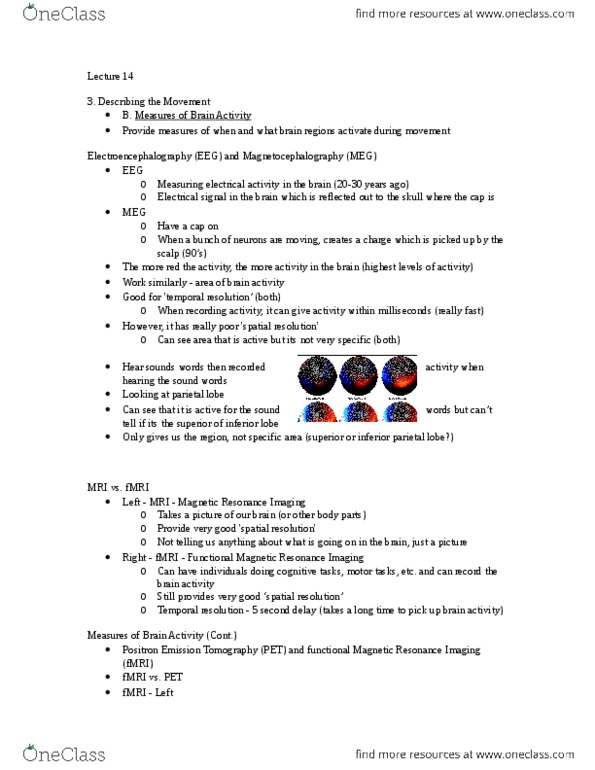
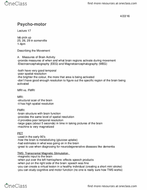
Friday, february 6, 2015: describing the movement. Provide measures of when and what brain regions activate during movement. Came about 35 or so years ago. Electrodes attached to head, electrical signal within brain gets reflected back to skull and electrodes pick up signal. Magnetic charge from neurons moving picked up by scalp. Picture: each map from same person the more red, the more activity there is in the brain. Both techniques provide us with location of brain activity. Good for temporal resolution (can give us brain activity within milliseconds - fast) However, it has poor spatial resolution (cid:523)can see area of brain that(cid:495)s active, but its not. E. g. , looking at parietal lobe can see that its active for sound words, but you can(cid:495)t tell whether its superior, inferior, etc. Just taking a picture not telling us whats going on in the brain fmri: functional magnetic resonance imagine. Can have individuals do different tasks (cognitive, cognitive-motor, etc. )