Biology 2382B Lecture Notes - Lecture 13: Red Blood Cell, Immunoglobulin Heavy Chain, Profilin

30 Mar 2016
School
Department
Course
Professor
Document Summary
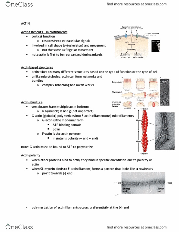
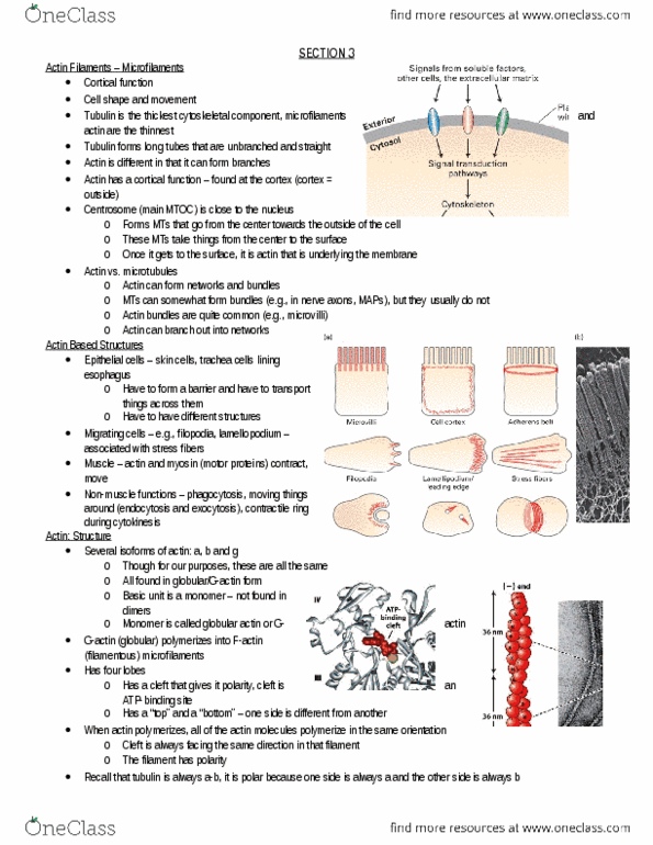
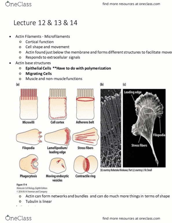
Most are cortical (work just under cell surface), whereas mts brings things to surface and hand them off to actin. G-actin monomer: globular shape with atp binding domain, which always polymerizes in the same orientation. Alpha (muscle), beta, and gamma isoforms, but all do the same thing. When decorated with myosin 1, arrow head pattern points to (-) end, and actin is stabilized. At cc, polymerization occurs preferentially at (+) end. Can form complex bundles and networks, whereas mts only form bundles. No nucleating factor = lag while nucleating factor is made, then elongation. Steady state: maintaining cc; move above cc and polymerize one monomer, bring you below cc where you depolymerize one monomer. Not functionally important because doesn"t happen in real life. Remove adp monomer, but need to add atp form (hydrolysis occurs after polymerization) (-) end cc: 0. 60 microm (+) end cc: 0. 12 microm.