Biology 2382B Lecture Notes - Lecture 3: Silver Stain, Peptide, Lipid Bilayer

22 Jan 2015
School
Department
Course
Professor
Document Summary
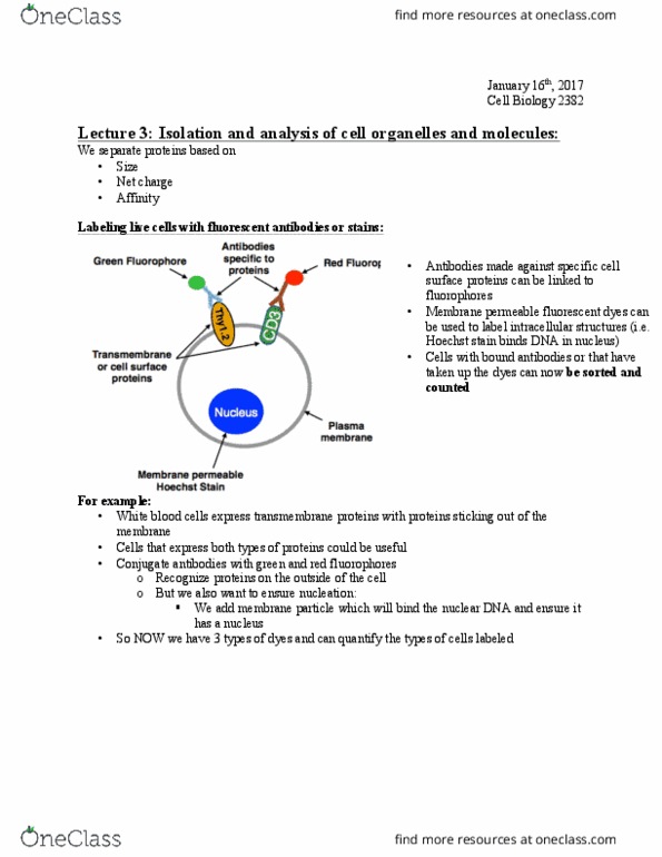
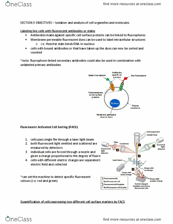
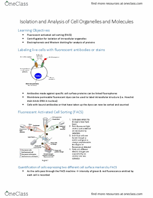
Live cells can be labeled with fluorescent dyes without killing the cell. Some cells contain transmembrane surface proteins which antibodies tagged with different fluorophores can attach to. Note that fluorophore-linked secondary antibodies could also be used in combination with unlabeled primary antibodies. In addition, membrane permeable fluorescent dyes can also be used to label intracellular structures such as hoechst stain that binds to dna in the nucleus. Cells with bound antibodies or that have taken up the dyes can now be sorted and counted. These cells then pass in a single file line through a laser light beam. As they continue in this line, both fluorescent light emitted and scattered are measured by detectors. As the individual cells exit, they are forced through a nozzle and given a charge proportional to the degree of florescence detected. Finally cells with different electric charges are separated by an electric field and collected.