Anatomy and Cell Biology 3319 Lecture 1: Heart-Structure & Function

9 Mar 2017
School
Department
Professor
Document Summary
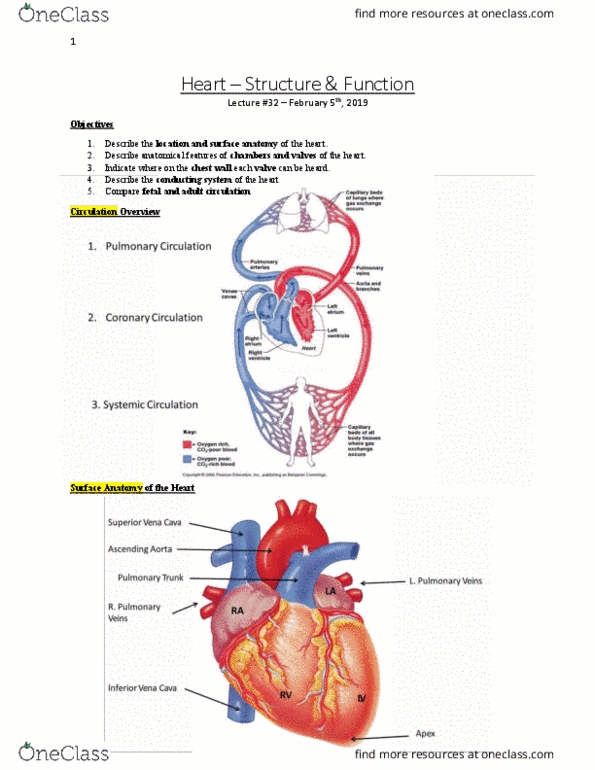
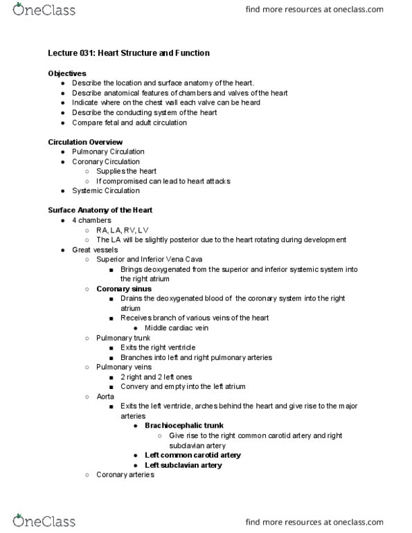
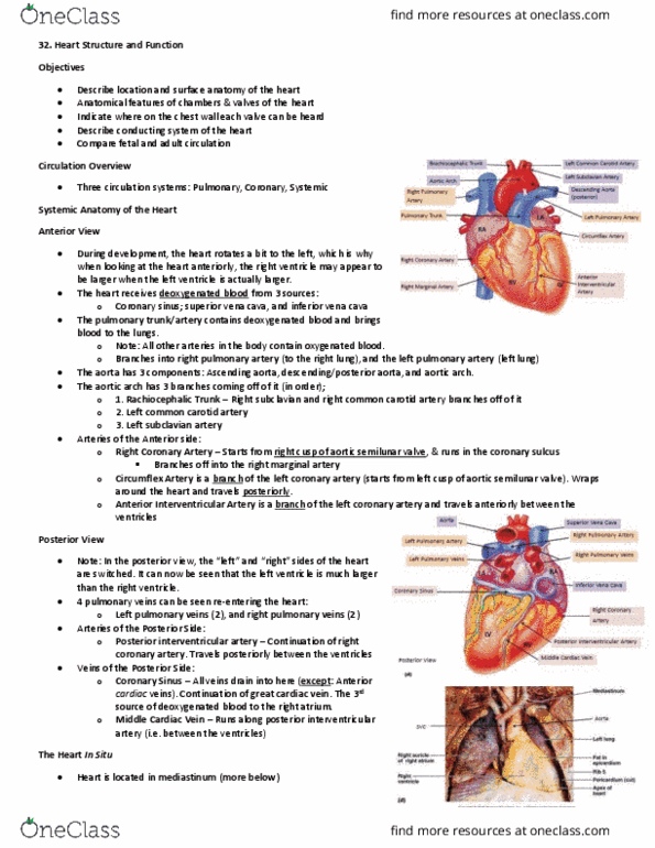
*during development the heart rotates abit to the left: deoxygenated blood going into ra: R/l pulmonary trunk going to r/l lung: oxygenated blood from lungs into heart: 2 pulmonary veins (r/l) ultimately flowing through la: blood pumped from. La to lv and moves into aorta (ascending, arch, descending) travels down to the pelvis: off of the arch there are 3 arteries (brachiocephalic, lc carotid, l subclaviann) Apex: tip of the heart, 5th intercostal space. Auricle: blind sac attached to the r/l atrium. Borders: right: right atrium (ra, left, superior: great vessels originate from here (aorta, pulmonary vessels) Corners: upper rc: 3rd right costal cartilage @ sternum, upper lc: 2nd left costal cartilage left of sternum, lower rc: 6th right costal cartilage @ sternum, lower lc: 5th intercostal space @ mid clavicular line. Mediastinum: middle portion of the chest where the heart is found . In the embryo it is a foramen ovalis.