Anatomy and Cell Biology 3319 Lecture Notes - Lecture 23: Superior Orbital Fissure, Condyloid Process, Zygomatic Bone

22 Nov 2016
School
Department
Professor

Nov 17 – Osteology of the Skull
Cranial cavity = inside of skull
Frontal bone – i fetus starts as to oes that fuse ad if do’t totall fuse get etopi suture hih looks like frature
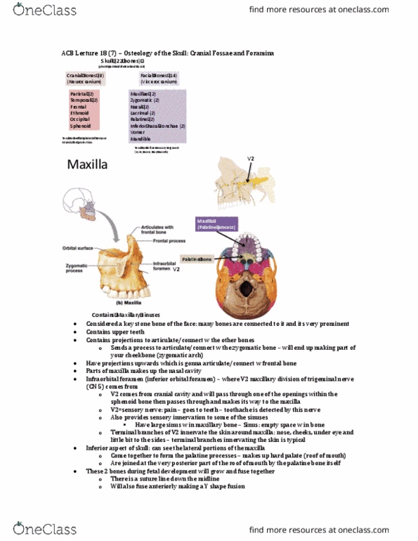
Maxilla – paired bone, forms inferior part of orbit
- If you take on maxillary bone and look from side, you can see it articulates with
zygomatic bone
- Part of maxilla that reaches out and touches with zygomatic bone = called
zygomatic process
- Frontal process – part of maxilla that reaches up that touches frontal bone
- Infraorbital foramen – where V2 of trigeminal nerve comes and innervates face
- Alveolar margin – sockets for teeth
- Part of oe a’t see i this ie – palatine plate – part of maxilla that
travels inward and forms part of roof of mouth (hard palate) (see last lecture inferior
view of skull)
- Soeties foratio of ailla ad palatie oe does’t happe ell
- The hard palate forms from two out growths that start at side and move
towards the midline (called palatine shelves) – if something wrong and two palatine
sheles do’t oe together ou get left lip
- Has flat part called squamous region
- Zygomatic process – forms zygomatic arch (cheekbone)
- Mastoid region towards back – bump – point of attachment for muscles for
moving skull
- Styloid process – point of attachment for muscles
- Most important = mandibular fossa – articulates with the mandibular condyle
of mandle
- Temporal mandibular joint (TMJ) every time you move your jaw
- 1st diagram looking at temporal bone from outside, second = inside
- petrous (rocky) ridge – divides inside of cranial cavity into middle and posterior
cranial fossa
- Frontal – yellow
- Zygomatic – blue
- Maxillary – purple
- Pink – part of sphenoid bone –
contributes to formation of superior
orbital fissure (crescent shaped hole) –
nerves travel here
- Orange – ethmoid
- Palatine bone – white (small)
- Green – lacrimal bone
- get punched in eye, fracture to
lower bone therefore soft part of lower eye socket comes out form orbit down into maxilarry sinus
- floor and medial wall are super thin – damage
Sphenoid – looking down into cranial cavity
- cant really see it from outside, but key bone from inside
- has body, small and large wing
- sella turcica (turkish saddle) –where pituitary gland will be nessled and protected
- has a lot of foramina where nerves will one through
o Optic canal where optic nerve will go through
o Superior orbital fissure – nerves that travel to extra ocular muscles
o In greater wing three more formina
▪ Rtundum (V2 trigeminal nerve, and V3), ovale, spinosum * know what
structures travel through these
find more resources at oneclass.com
find more resources at oneclass.com
Document Summary
Frontal bone i(cid:374) fetus starts as t(cid:449)o (cid:271)o(cid:374)es that fuse a(cid:374)d if do(cid:374)"t totall(cid:455) fuse get (cid:373)etopi(cid:272) suture (cid:449)hi(cid:272)h looks like fra(cid:272)ture. Maxilla paired bone, forms inferior part of orbit. If you take on maxillary bone and look from side, you can see it articulates with. Part of maxilla that reaches out and touches with zygomatic bone = called. Frontal process part of maxilla that reaches up that touches frontal bone. Infraorbital foramen where v2 of trigeminal nerve comes and innervates face. So(cid:373)eti(cid:373)es for(cid:373)atio(cid:374) of (cid:373)a(cid:454)illa a(cid:374)d palati(cid:374)e (cid:271)o(cid:374)e does(cid:374)"t happe(cid:374) (cid:449)ell. The hard palate forms from two out growths that start at side and move. Mastoid region towards back bump point of attachment for muscles for. Styloid process point of attachment for muscles. Most important = mandibular fossa articulates with the mandibular condyle moving skull of mandle. Temporal mandibular joint (tmj) every time you move your jaw.