Anatomy and Cell Biology 3309 Lecture Notes - Lecture 17: Muscular Layer, Muscularis Mucosae, Stratified Squamous Epithelium

22 May 2018
School
Department
Professor

Histology 3309
Esophagus & Stomach
Learning Objectives
1. Explain the general organization of the GI tract
2. Name the different layers and their main functions
3. Distinguish upper, intermediate and lower sections of the esophagus
according to muscle type in the muscularis externa
4. Differentiate between esophagus and stomach according to surface epithelium
5. Characterize the stomach mucosa as having pits and glands
6. Describe 6 different cell types in the lining epithelium of the stomach and name their functions
7. Describe the location and function of auerbach’s and meissner’s nerve plexus
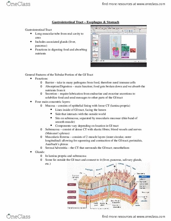
Layers and functions of the GI Tract
- 4 main concentric layers:
1. mucosa
▪ mucosa is the inner lining of the tubular GI tract
▪ consists of an epithelium (this interacts with the food that comes in)
▪ located on the lamina propria (loost CT)
2. submucosa
▪ dense irregular CT
▪ carries into the mucosa things like nerves, blood vessels etc
3. muscularis externa
▪ the tube is surrounded by this layer of smooth muscle
▪ allows the gut to move the food along
4. adventitia or serosa
▪ DICT or mesothelium
▪ So we have a dense connective tissue covering called the adventita
▪ But in certain parts of the GI tract is hanging in the body cavity and in those
places that is hanging, instead of having an adventitia, we have a layer of
metholium (lining of body cavity) which is aka serosa
- Functions of the GI tract:
- Barrier to the outside world
o The gut associated lymphoid tissue (GALT) lives in the mucosa ro the submucosa
- Absorption/digestion – specific parts o the mucosa are differentiated to do that
- Secretion: endo- and exocrine
- In order to facilitate absorption/digestion, we have glands either in the mucosa or the
submucosa to provide lubrication to move things and liquidify the food content
find more resources at oneclass.com
find more resources at oneclass.com

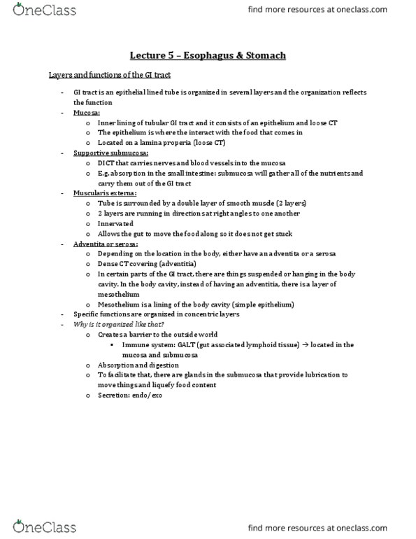
The General Organization of the GI Tract
- Learn this slide – this is your roadmap of the GI tract
- Along the length, you see the different regions that the food passes through in order to nourish
us →Esophagus and stomach to Small and large intestine
- Submucosa and the muscularis externa look very similar along the length of the GI tract
- Mucosa is where there is a lot of changes that help distinguish the different subparts of the tract
Esophagus
- Designed to deliver the food into the stomach
- Epithelium lined tube that has a mucosa, submucosa (not really shown here)
- The muscle layer in the muscularis externa changes – the type of
muscle you find depends on location
- The upper third of the esophagus has a muscularis externa composed
of mainly striated muscle (voluntary muscle) →meaning that we are
able to actively regurgitate stuff
- There are species which their life depends on that (birds that feed their
young, actively regurgitate food)
- At the lower end, towards the stomach, its predominantly smooth
muscle
- Both types of muscle start mixing in the middle
- So if you get a section of esophagus and can tell where the section is
roughly taken from by looking at the type of muscle found
- this is a low mag overview of the esophagus
- at this magnification, you cant really determine that this is smooth muscle but here we have 2
layers of smooth muscle
o the inner layer runs circumferentially – the contraction of the inner layer changes the
size of the lumen
o the outer layer runs longitudinally – the contraction of the outer layer will
compartmentalize segments of the tube
o together, the contraction will be abel to move food substances along
- the mucose consists of an epithelium
- both mucosa and muscularis externa is separated by a dense connective tissue, submucosa
- in certain regions, you find within the submucosa, mucous secreting glands that are designed to
lubricate the surface of the mucosa (the inner surface)
find more resources at oneclass.com
find more resources at oneclass.com

- the lumen of the esophagus is always highly folded – irregular
o this allows diff consistencies of food to be accommodated
o if we swallow big pieces, the mucosa can extend and is adaptable to the size or the
consistency of the food we take in
- this is higher magnification of the esophagus
- the esophagus has an epithelium that is stratified squamous non-keratinized (very similar to the
oral cavity)
- this type of epithelium is mainly found in 2 places in the body:
o esophagus
o vagina
- this epithelium reflects the function of protection and wear and tear
- we also have a lamina propria
o very cellular underneath the loose connective tissue
- together with the lamina propria and the epithelium, we make the mucosa
- muscularis mucosae:
o component of the mucosa
o band of smooth muscle that separates the lamina propria from the submucosa
o not that visible in this slide – but very visible in the small and large intestine
- the submucosa is dense connective tissue, it has ducts that carry mucous from the submucosal
gland to the surface
- note: this is a PAS stained slide – the mucous in the gland appears red
- in this image the esophagus has stratified squamous epithelium, but when the esophagus
merges into the stomach, the epithelium changes to a simple columnar epithelium
find more resources at oneclass.com
find more resources at oneclass.com
Document Summary
Barrier to the outside world: the gut associated lymphoid tissue (galt) lives in the mucosa ro the submucosa. Absorption/digestion specific parts o the mucosa are differentiated to do that. In order to facilitate absorption/digestion, we have glands either in the mucosa or the submucosa to provide lubrication to move things and liquidify the food content. Learn this slide this is your roadmap of the gi tract. Along the length, you see the different regions that the food passes through in order to nourish us esophagus and stomach to small and large intestine. Submucosa and the muscularis externa look very similar along the length of the gi tract. Mucosa is where there is a lot of changes that help distinguish the different subparts of the tract. Designed to deliver the food into the stomach. Epithelium lined tube that has a mucosa, submucosa (not really shown here) The muscle layer in the muscularis externa changes the type of.