Anatomy and Cell Biology 3309 Lecture Notes - Lecture 22: Outer Plexiform Layer, Inner Nuclear Layer, Outer Nuclear Layer

15 May 2018
School
Department
Professor

Lecture 22 – The Retina
The eye
- Retina is part of the CNS
o Information processing already occurs here
o Detection of movement, orientation of edges, contrast enhancement is done in the
retina before the information reaches the rest of the brain
- Optic nerve is continuous with the retina (most inner tunic of the eye)
- Retina =
o Neural retina (the tissue containing photoreceptors used to see)
o Retinal pigment epithelium (outer layer to the neural retina)
o Continuous with two epithelial layers that cover the ciliary body and iris
▪ OUTER: pigmented and continuous with the retinal pigment epithelium
- Histological slide of the retina
- Retina is very layered and nicely organized
- Layer in the box is the retinal pigment epithelium (outer layer)
- Retinal pigment epithelium: one layer of epithelial cells that borders the choroid and there
is a membrane in between
- Beneath the retinal pigment epithelium is the choroid
- Layers with the cell bodies are called nuclear layers
- There are layers with no cell bodies called plexiform layers - neurophil (dendrites,
synpases) → this is where neurons make contact
- Light comes from the top of the image (where the inner eye is)
- The photoreceptors are the long basophilic nuclei with their outer segment
o This is where phototransducton takes place
o Light must travel throughout the entire retina to hit the outer segment of the
photoreceptor to then induce a neuronal signal
- Red dots are blood vessels
- There are blood vessels in the inner portion of the retina but not in the outer portion
find more resources at oneclass.com
find more resources at oneclass.com

- There is no vascularization in the outer portions of the retina and they are provided with
nutrients through the choroid and retinal pigment epithelium to the outer segment of the
photoreceptors
The retina
- From outside → inside:
o 1. Pigment epithelial cells (most outer layer)
o 2. Photoreceptors (outer nuclear layer)
o 3. Pexiform layer (outer plexiform where there are synpases of photoreceptors to
the next cells)
o 4. Inner nuclear layer (horizontal, bipolar, amacrine cells)
o 5. Inner plexiform layers (no cell bodies)
o 6. Ganglion cell layer (cell bodies of retinal ganglion cells) – only cells that fire APs in
the retina, other cells are neurons but they do not fire APs, only graded APs
o 7. Ganglion cells fire APs and axons travel through nerve fiber layer to the optic disk
and merges into the optic nerve
o 8. Axons of ganglion cells project into the brain
find more resources at oneclass.com
find more resources at oneclass.com

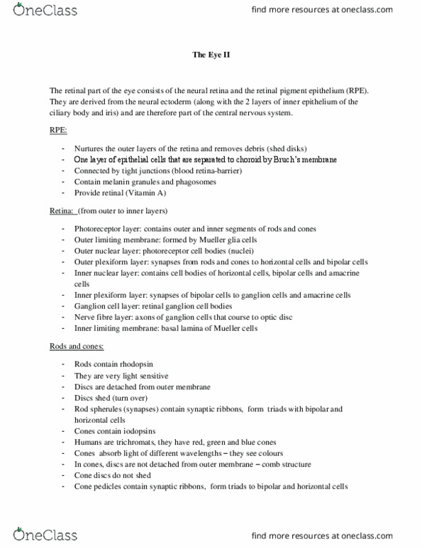
The retinal pigment epithelium (RPE)
- Most outer layer
- Border the choroid
Epithelial cells have different functions:
- 1. Pigmented: contain melanocytes = also absorb light that was not absorbed at the
photoreceptor cells
o The light be absorbed because otherwise it would enter the brain/skull
o Retinal pigment epithelial cells contain melanocytes for light absorption
o Some animals have retinal cells that reflect light instead of absorbing it to see at
night = their eyes glow = another chance to hit a photoreceptor cell
▪ Not all light is absorbed in the photoreceptor cell and light is emitted = light
shines at night in nocturnal animals
o In humans and not night active, the retinal pigment epithelial cells absorb the light
- 2. Transport nutrients and ions
o Photoreceptor cells are neurons and they must maintain homeostasis in terms of ion
concentration around the photoreceptor cells
- 3. Glial cell function
o Provide nutrients that the photoreceptors cells need
o Buffer potassium ions
- 4. Visual cycle
o There is a pigment in the outer photoreceptor segments (photosensitive pigment)
o Pigment must be constantly recycled because when a photon hits it, it undergoes a
conformational change and it leads to the opening of an ion channel
o Retinal pigment epithelial cells recycle the photopigments
- 5. Microglia cell function:
o Phagocyte debris from the retina
find more resources at oneclass.com
find more resources at oneclass.com
Document Summary
Information processing already occurs here: detection of movement, orientation of edges, contrast enhancement is done in the retina before the information reaches the rest of the brain. Optic nerve is continuous with the retina (most inner tunic of the eye) Retina is very layered and nicely organized. Layer in the box is the retinal pigment epithelium (outer layer) Retinal pigment epithelium: one layer of epithelial cells that borders the choroid and there is a membrane in between. Beneath the retinal pigment epithelium is the choroid. Layers with the cell bodies are called nuclear layers. There are layers with no cell bodies called plexiform layers - neurophil (dendrites, synpases) this is where neurons make contact. Light comes from the top of the image (where the inner eye is) There are blood vessels in the inner portion of the retina but not in the outer portion.