Anatomy and Cell Biology 3309 Lecture Notes - Lecture 21: Retinal Pigment Epithelium, Neural Tube, Retina

15 May 2018
School
Department
Professor

Lecture 21 – The Eye I
- Sensory organ
- Part of the eye is part of the brain
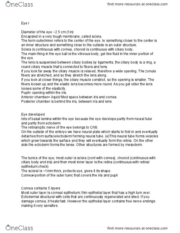
- Human eye diameter is 2.5 cm
- Tunics are layers of the eye
o Sclera: outer layer
o Choroid
o Retina: most inner layer
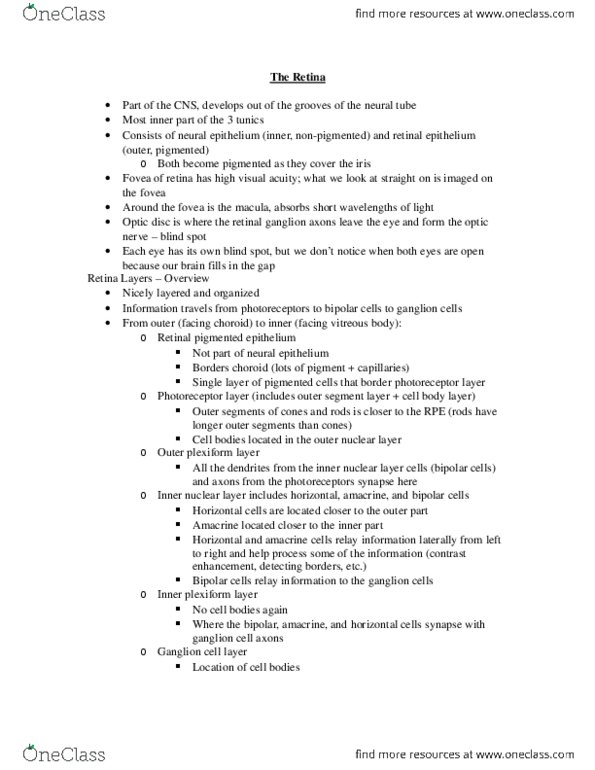
- RETINA: retina + retinal pigment epithelium
o Consists of neural retina and retinal pigment epithelium
o Fovea and macula are regions of the retina
- Viterous body: lumen/interior of the body
- Structures within the eye:
o INNER: towards the center of the eye
o OUTER: towards the outside of the eye
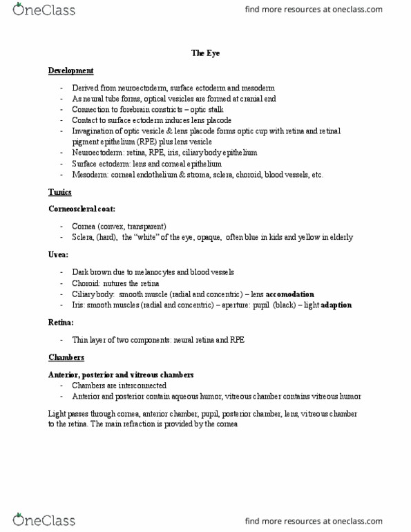
Development
- Structure of the eyes look similar in the animal kingdom but they develop differently
- Neural tube develops from the embryo which gives rise to the spinal cord and brain
o Folds from the ectoderm of the embryo and neural tube detaches from the outer
layer of the embryo
o At the cranial end of the fetus, the neural tube becomes the brain
- Head end of the fetus and neural tube form vesicles that then become the brain
- Vesicles form other vesicles that will later become the retina
- Grow out of the neural tube towards the surface of the embryo at the head end
- Outer area become the retinal pigment epithelium (green) and the pink is the future neural
retina (real retina used for vision)
- Vesicle grows out of the neural tube towards the surface and starts to invaginate
- At the same time, the outer surface starts to form the lens and invaginates as well
find more resources at oneclass.com
find more resources at oneclass.com

- Outer layer (ectoderm) becomes the length
- What grew out of the neural tube becomes like a cup
o There are two layers: green and pink that lie on top of each other
o Becomes retinal pigment epithelium and retina
- It is still connected to the neural tube (the part that will become the brain) by a stalk that
becomes the optic nerve
- Retina, retinal pigment epithelium with the optic nerve directly connect to the rest of the
brain (THEY ARE PART OF THE CNS)
o Basically parts of the brain grew out of the brain
- There is bbb in the CNS and in the retina (blood retina barrier)
- Other parts of the eye are from the ectoderm (lens, epithelium of cornea)
- Other parts of the eye are derived from mesenchymal structures
- Retina is an easily accessible part of the brain (IT IS A PART OF THE BRAIN)
Tunics of the eye
- Tunics surround the eye
- SCLERA: outermost layer
o First cut into the eye is very tough because it surrounding by sclera
o Very tough layer surrounding the eye
o Keeps the eye in shape
o Continuous with the dura matter
o Continuous with the cornea at the front of the eye
- UVEA:
o Contains choroid (layer containing blood vessels), ciliary body and iris
o IRIS + CILIARY BODY + CHOROID = FORM THE INNER LAYER
o Choroid and arachnoid have similar structures and function
- RETINA:
o Most inner layer that grows out of the brain and forms the optic nerve
o Retina contains retinal pigment epithelium and neural retina (two layers)
find more resources at oneclass.com
find more resources at oneclass.com

o Continues as a double layer of epithelial cells at the inner edge of ciliary body and
the iris
o Area covering the ciliary body and iris are still part of the retina (derived from CNS
structure)
o Neural retina is the part of the eye with which we see
- Corneoscleral coat
o Cornea and sclera, ~1mm thick, protects and shapes the eye
- Uvea
o Choroid, ciliary body and iris, vascular layer
- Retina
o Retinal Pigment Epithelium (RPE) and neural retina
The choroid
- Bruch’s membrane
o Choroid is separated from the retina by Bruch’s membrane basement membrane
o Separates retinal pigment epithelium from the choroid
o Inner most
o Basal lamina of opposing RPE and endothelial cells
o After Brunch’s membrane is the choroid itself
- Choroid contains two different layers: choriocapillaris + choroid stroma
o Both layers contain melanocytes (pigmented)
o Light not absorbed by retinal pigment is absorbed by the choroid and it makes sure
it does not go any further
- Choriocapillaris
o Inner portion of the choroid
o Fenestrated capillaries
o Supplies nutrients to the outer retina
o It is difficult to get nutrients to the outer part of the retina because it is not
vascularized in humans (no blood vessels)
o Nutrients are supplied from the capillaries in the choroid
find more resources at oneclass.com
find more resources at oneclass.com
Document Summary
Part of the eye is part of the brain. Tunics are layers of the eye: sclera: outer layer, choroid, retina: most inner layer. Retina: retina + retinal pigment epithelium: consists of neural retina and retinal pigment epithelium, fovea and macula are regions of the retina. Inner: towards the center of the eye: outer: towards the outside of the eye. Structure of the eyes look similar in the animal kingdom but they develop differently. Head end of the fetus and neural tube form vesicles that then become the brain. Vesicles form other vesicles that will later become the retina. Grow out of the neural tube towards the surface of the embryo at the head end. Outer area become the retinal pigment epithelium (green) and the pink is the future neural retina (real retina used for vision) Vesicle grows out of the neural tube towards the surface and starts to invaginate.