Anatomy and Cell Biology 3309 Lecture Notes - Lecture 19: Fallopian Tube, Oviduct, Meiosis

15 May 2018
School
Department
Professor

Lecture 19 – Oviduct, Uterus, Vagina
- Ovulation occurs from LH surge which induces completion of meiosis I
o Before ovulation, there is a primary oocyte that is arrested at prophase if meiosis I
o Oocyte enter meiosis II and arrests in metaphase of meiosis II, becoming a
secondary oocyte producing a polar body
o Secondary oocyte is released into the body cavity and does not enter fallopian tube
directly
o Fallopian tube lies close to the ovary
o Ovary is within the lumen of the fallopian tube
- Oocyte may encounter spermatozoa in fallopian tube
- Fallopian tube is where female and male gametes meet and meiosis II completes
o = have two nuclei fuse to form a zygote
- Early cell division takes place and at the blastocyst stage of development, zygote finds its
way into the uterus where it implants into the wall
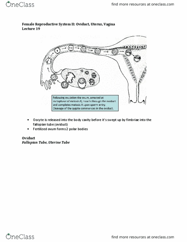
- Following ovulation the ovum, arrested at metaphase of meiosis II, travels through the
oviduct and completes meiosis II upon sperm entry
- Cleavage of the zygote commences in the oviduct
Oviduct – fallopian tube, uterine tube
- Fallopian tube is an open ended epithelial lined tube that lies closely adjacent to the surface
of the ovary
- Fallopian tube has finger like projections at its outer rim where it actually opens → this
forms a brush-like structure
- There are smooth muscle cells within finger-like projections and during ovulation, the
fimbrae sweep over the surface of the ovary to make sure they catch ovulating oocytes and
guide them into the lumen of the fallopian tube
- Fallopian tube is divided anatomically and structurally into three sections
- AMPULA: longest section
find more resources at oneclass.com
find more resources at oneclass.com

o Has one lumen and it curves around a little bit
o Histological section shows two cross sections of the lumen of the tube
o Connective tissue and smooth muscle are on the outside
- ISTHMUS: tube narrows to from the ampulla
o Penetrates through the wall of the uterus
- INTRAMURAL SEGMENT: passageway that travels through the wall of uterus
o Same thickness as the wall of the uterus
Ampulla of oviduct
- Lumen is obstructed by folds
- Mucosa forms branching folds that almost cover the whole lumen
- Epithelium and loose CT lines it
- If oocyte/zygote are travelling through the oviduct, it will always be in contract with the
surface and this allows for proper nourishment of the zygote and transport along its lumen
- Contact with the wall of the oviduct is important
- Even at low magnification, it is a prominent view → KNOW HOW TO RECOGNIZE
find more resources at oneclass.com
find more resources at oneclass.com

- The ovum is in intimate contact with the epithelium. Non-ciliated cells secrete fluid. Ciliated
cells generate a flow toward the uterus
- Histological image is one of the folds
- Below the cells is loose CT (lamina propria)
- Epithelium overlying the CT is simple columnar even though nuclei are at different locations
TWO CELL TYPES:
- 1. Non ciliated cells have no surface modifications and produce a fluid within the fallopian
tube that contains a lot of nutrients
- 2. Ciliated cells produce a flow of the fluid towards the uterus
o They are constantly beating and fluid flow moves the oocyte towards the uterus
o Spermatozoa must swim against the slow in order to reach the oocyte
- The number of ciliated cells changes depending on the hormonal situation during the
menstrual cycle
- After ovulation when progesterone levels rise, higher levels of progesterone will induce
differentiation of ciliated cells and increase beating to ensure any oocyte that has been
released during ovulation will be carried along
- EM image:
o Shows how space between adjacent epithelial cells (channels) through which oocyte
passes is full of ciliary extensions
o There is a constant sweeping motion
o Basal bodies are present = cilia are projecting into the lumen of the fallopian tube
- Hormonal changes during the menstrual cycle change the differentiation state of the
epithelial cells within the oviduct
find more resources at oneclass.com
find more resources at oneclass.com
Document Summary
Oocyte may encounter spermatozoa in fallopian tube. Fallopian tube is where female and male gametes meet and meiosis ii completes: = have two nuclei fuse to form a zygote. Early cell division takes place and at the blastocyst stage of development, zygote finds its way into the uterus where it implants into the wall. Following ovulation the ovum, arrested at metaphase of meiosis ii, travels through the oviduct and completes meiosis ii upon sperm entry. Cleavage of the zygote commences in the oviduct. Fallopian tube is an open ended epithelial lined tube that lies closely adjacent to the surface of the ovary. Fallopian tube has finger like projections at its outer rim where it actually opens this forms a brush-like structure. There are smooth muscle cells within finger-like projections and during ovulation, the fimbrae sweep over the surface of the ovary to make sure they catch ovulating oocytes and guide them into the lumen of the fallopian tube.