Anatomy and Cell Biology 3309 Lecture Notes - Lecture 12: Hyaline Cartilage, Bronchus, Trachea

15 May 2018
School
Department
Professor

Lecture 12 – Lung
Review
Epithelium
Cartilage
Smooth muscle
Other
Trachea
Respiratory
epithelium
Present in C-shaped
rings (hyaline
cartilage)
Trachealis muscle
(controls diameter of
trachea during
inhalation and
exhalation and allows
food to go down
esophagus)
• Submucosal glands
• Mucus secreting
glands
• Contains lymphatic
tissue (BALT)
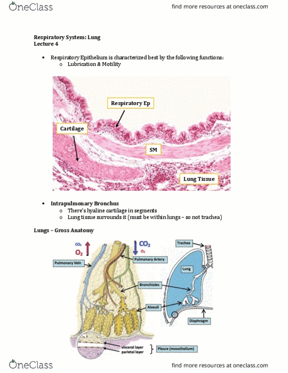
Bronchi
(intra-
pulmonary)
Respiratory in
trachea → simple
columnar in the
bronchioles
• Further from
trachea it
becomes low
columnar
Discontinuous
cartilage plates
(hyaline cartilage)
• Further you move
from trachea into
intrapulmonary
bronchus, the less
cartilage is
involved
Layer between lamina
propria and submucosa
(goes all the way
around –
circumferential)
• Contains
submucosal glands
(mucus secreting)
• BALT
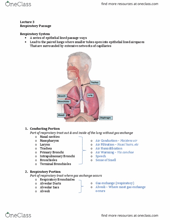
Respiratory tract
- Lose mucous secreting glands and BALT as you move down the respiratory tract
The lung
- Trachea splits into two extrapulmonary bronchi (outside of the lung bronchi)
find more resources at oneclass.com
find more resources at oneclass.com

- When bronchi enter the lungs they are now intrapulmonary bronchi
- When you breathe, rib cage moves out and diaphragm moves down to make the space
inside the rib cage larger
- Lungs are adhered to the rib cage via pleura and they follow the rib cage
- Diaphragm muscle and lung tissue are lined by a single layer of squamous shaped epithelial
cells that make up the pleura
o Referred to as mesothelium with a visceral and parietal layer
o Visceral layer of the pleura is on the outside
o Parietal pleura is adhered to diaphragm muscle and inside of the rib cage
o Two layers are continuous with each other
- Most common form of cancer of the lungs is mesothelioma
- Lungs have vessels (purpose of breathing is to oxygenate blood)
o Pulmonary artery: carries deoxygenated blood
▪ Only artery that carries deoxygenated blood (comes from the heart and goes
to the lung)
o Pulmonary vein: carries oxygenated blood
- Intrapulmonary bronchus → terminal bronchiole → respiratory bronchiole → alveoli
- Artery and vein make a network of capillaries around the alveoli
Terminal bronchioles
- Smaller airways → < 1 mm
- No cartilage
- No secretion of mucous
- Lots of smooth muscle
- Simple cuboidal epithelium
- Clara cells present
find more resources at oneclass.com
find more resources at oneclass.com

- Intrapulmonary bronchus → terminal bronchioles
o NOT BRONCHI → lose a few things
o Lose cartilage and mucous glands
o Increase smooth muscle and elastin as you go further down into the bronchioles
- Characteristics:
o All bronchioles do not have cartilage
o Bands of smooth muscle that run circumferentially around the bronchiole
o Elastin fibers are present in bronchioles and alveoli
o Moving into the bronchioles, there is a transition into a simple cuboidal epithelium
o Epithelium has clara cells
- ???: smooth muscle
o Makes circumferential ring around terminal bronchiole
- Center is the lumen of the bronchiole
- Simple cuboidal epithelium: one layer of cells that are as short as they are wide
- Alveolus: white space
o Around the bronchiole, there is no direct connection to the alveolus
o Vs. intrapulmonary bronchi have alveolar tissue around as well because they are
within lung tissue
o Vs. extrapulmonary bronchi do not have alveoli around
find more resources at oneclass.com
find more resources at oneclass.com
Document Summary
Respiratory in trachea simple columnar in the bronchioles: further from trachea it becomes low columnar. Discontinuous cartilage plates (hyaline cartilage: further you move from trachea into intrapulmonary bronchus, the less cartilage is involved. Trachealis muscle (controls diameter of trachea during inhalation and exhalation and allows food to go down esophagus) Layer between lamina propria and submucosa (goes all the way around circumferential: submucosal glands, mucus secreting glands, contains lymphatic tissue (balt, contains submucosal glands (mucus secreting, balt. Lose mucous secreting glands and balt as you move down the respiratory tract. Trachea splits into two extrapulmonary bronchi (outside of the lung bronchi) When bronchi enter the lungs they are now intrapulmonary bronchi. When you breathe, rib cage moves out and diaphragm moves down to make the space inside the rib cage larger. Lungs are adhered to the rib cage via pleura and they follow the rib cage. Most common form of cancer of the lungs is mesothelioma.