PSL300H1 Lecture Notes - Lecture 10: Melanin, Rhodopsin

Document Summary
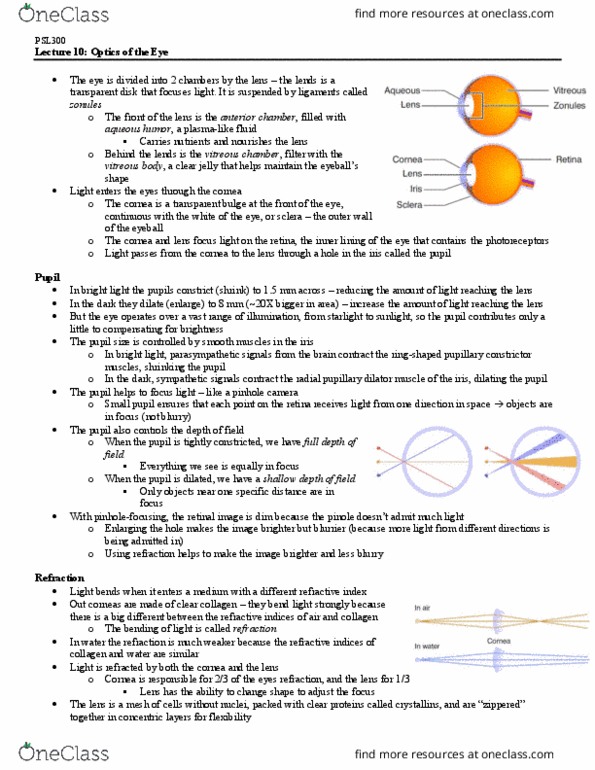
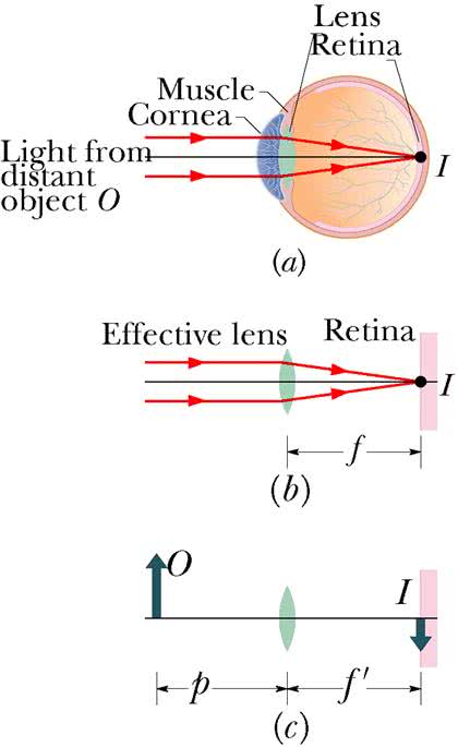
Visual system takes up a lot of space in the brain. Images are turned backwards and upside down on retina. Camera model pupil as aperture, lens, retina as film. Pin-hole pupil entire depth of field perfectly focused on retina for all objects in the field (whether far or near: one ray of light from every direction enters the eyeball. Dilate pupil require lens to refocus divergent beam of light. October 01, 2007: no hindrance to incoming light be other neurons, maximal visual acuity can detect fine detail. Blood vessels and axons enter/exit retina via blind spot . Pit in retina region of highest visual acuity small photoreceptors (cones) packed in high density blood vessels and axons encircle this area they do not cross it. No light membrane depolarized (-40 mv: cgmp is abundant in dark conditions and opens cgmp-gated na+ channels. 10 eyeball and retina o vesicles released constantly.