BIOB33H3 Lecture Notes - Lecture 1: Hyaline, Osteon, Synovial Joint


Document Summary
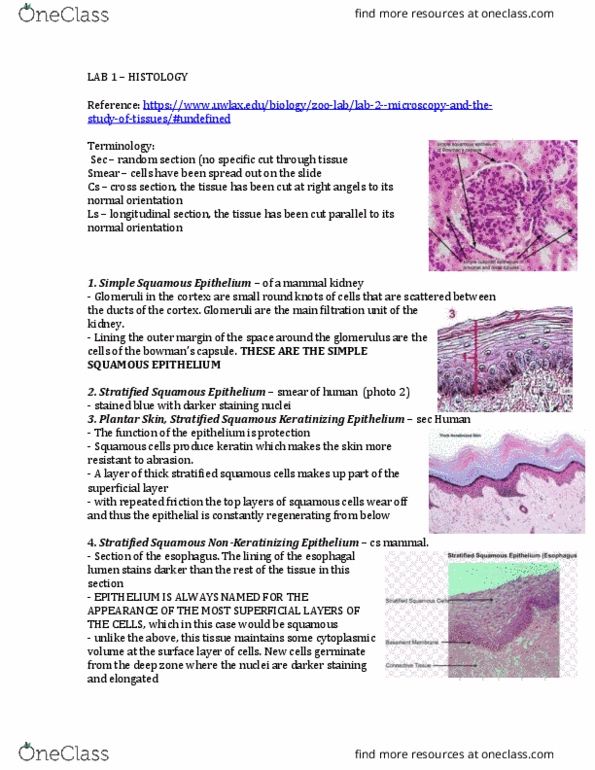
Then find the cells/structures referred to in the paragraph. If you have time, sketch a small diagram of what you see. First distinguish the cortex of the kidney from the medulla (see diagram below). They are small round knots of cells that are scattered between the ducts of the cortex. Glomeruli are the main filitration unit of the kidney. Lining the outer margin of the space around the glomerulus are the cells of bowman"s capsule. You can also see simple squamous epithelium forming the walls of the thin segments of the loops of henle in the medulla of the kidney. In this smear squamous epithelial cells are spread widely (kind of like spreading pieces of a jigsaw puzzle around). Note the pancake-like appearance of these cells, stained blue, with darker staining nuclei. You can not really appreciate the stratified nature of these cells from this section. For this, see the plantar skin" slide: plantar skin, stratified squamous keratinizing epithelium, sec.