BIOA01H3 Lecture Notes - Lecture 13: Microsoft Onenote, Optical Microscope, Green Fluorescent Protein

7 May 2015
School
Department
Course
Professor


18
BIOA01H3 Full Course Notes
Verified Note
18 documents
Document Summary
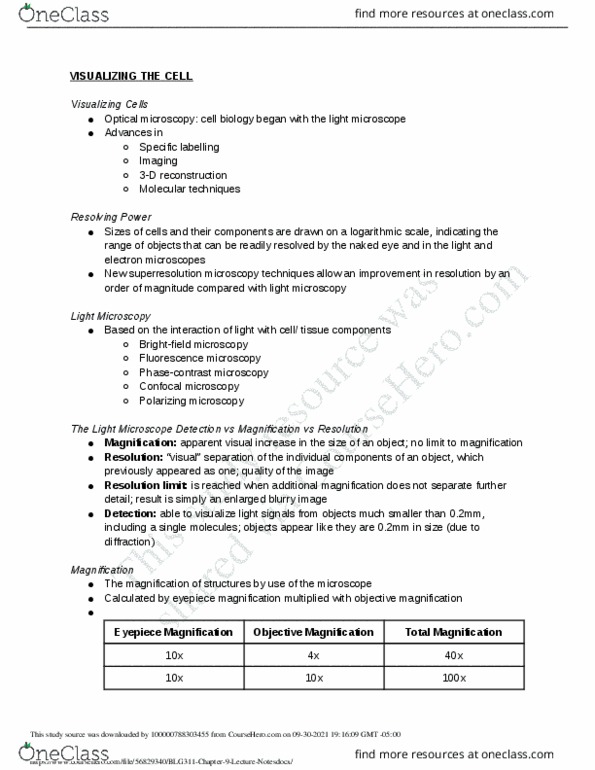
Looked at very simple organisms and detail their structure. Slow progression until the 1980s and then there was an explosion in knowledge of microscopy. Spatial resolution of biological imaging techniques https://onedrive. live. com/edit. aspx/documents/biology?cid=4612573e81e7633b&id=documents&wd=target%28september. one%7caa4f95b0-28e4 1/10. Microscopy techniques span across a variety of sizes. Electron microscopy has been around since turn of century and is really old compared to others. What they have in common: they all use a fluorescence in the excitation of the molecule of light in order to see structures. Light energy comes in packets (photons) that have wavelike characteristics. Photons contain a fixed amount of energy that is inversely related to its wavelength. So short wavelengths = high energy, long wavelengths, low energy. Photons are the ones that strike objects and then you see them. Visible light (white light) is 400nm to 700nm. In physics, they call ultraviolet and infrared light as well.