PHS 3342 Lecture Notes - Lecture 12: Bronchus, Respiratory Tract, Bronchiole

28 Apr 2018
School
Department
Course
Professor

March 6, 2018
Respiratory Mechanisms
Structure and Function
The entire process of respiration is based on diffusion of gases down their concentration gradients
To facilitate transport of oxygen and carbon dioxide, we use:
-Bulk flow of air in and out of the lungs
-Large surface area for gas exchange (alveoli)
-A closed circulatory system
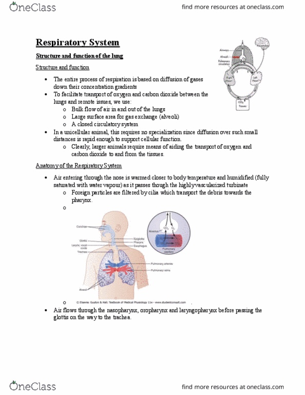
Anatomy of the Respiratory System
Lungs:
-Right lung: 3 lobes
-Left lung: 2 lobes
-Mediastinum: area between the two lungs where the heart resides
•Supported by the sternum/chest wall and the spine
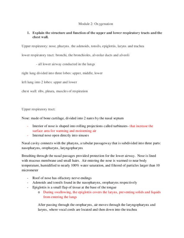
Passage of air:
-Air entering through the nose is warmed closer to body temperature and humidified (fully warmed with water
vapour) as it passes through the highly vascular turbinates
•Helped by a great number of ridges (nasal chonchae aka turbinates) inside the nose - increases blood flow and
heat available
-Foreign particles are filtered by cilia which transport the debris towards the pharynx
-Air flows through the nasopharynx, oropharynx, and laryngopharynx before passing the glottis on the way to the
trachea
-Air then flows down the trachea and divides into the right or left main bronchus (primary bronchi)
-The main bronchi divide into lobar bronchi (secondary), then segmental (tertiary) bronchi and again eventually to
terminal bronchioles, respiratory bronchioles, alveolar ducts, and alveoli
Conduction zone: responsible for bulk air flow
-Trachea
•Surrounded by rings of cartilage up until the end of the bronchi - cartilage helps prevent the airway from
collapsing
-Bronchi: right bronchus is more vertical than the left
-Bronchioles
1

March 6, 2018
Respiratory zone: responsible for gas exchange
-Respiratory bronchioles
-Alveolar ducts
-Alveoli: at the alveolar level, total lung surface area is so high that air flow becomes very slow (much like blood
velocity slows down in the capillaries)
•Air flow is essentially replaced by diffusion, explaining why this zone is known as the respiratory zone
Mechanisms of Breathing: Lung and Chest Cavity
Lung is surrounded by two pleura:
-Visceral pleura is directly in contact with the lung
-As the lung expands, it pushes the visceral pleura towards the parietal pleura
•NB: still don’t come in contact - there will still be a small gap between the two pleura (intrapleural space filled
with thin film of liquid)
-Pleura cause the lung to be virtually stuck to the chest wall - causes the lungs to expand when the chest wall
expands
Lung Volumes and Capacities
Tidal volume (TV): amount of air coming in and out at rest
-*TV = 500 mL
Inspiratory reserve volume (IRV): amount of air that can be forcibly inhaled (maximal inhalation)
Expiratory reserve volume (ERV): amount of air that can be forcibly exhaled (maximal exhalation)
Residual volume (RV): amount of air left in the lungs after forced maximal exhalation
-NB: can’t be measured using spirometry - only measures the amount of air that comes out, but can’t measure the
amount that’s left
Functional residual capacity (FRC): amount of lungs left in the air after regular exhalation (at rest)
-FRC = ERV + RV
-NB: can’t be measured using spirometry - RV can’t be measured
Vital capacity (VC): amount of air moving in and out from maximal exhalation to maximal inhalation
-VC = TV + IRV + ERV
Total lung capacity: maximal volume of air that can be contained in the lungs
-TLC = VC + RV
-NB: can’t be measured using spirometry - RV can’t be measured
2

March 6, 2018
NB: in general, capacities are sum of other volumes
Measuring Lung Volumes
Spirometry: measure the amount of air moving in and out of the lungs
-Person breathes through a tube (nose is plugged)
-Tube is connected to a can that is reversed in water
-Breathing in makes the can fall
•Can is connected to a pen that is tracing on paper - cause pen to move upwards and draws
-Exhalation makes the can rise - pen moves downwards on the paper
Helium dilution method: used to measure residual volume
-Person breathes through a tube
-Tube has an initial volume and concentration of helium
-Amount of helium at the beginning will represent the amount of helium available at the end
•Closed system - no helium escapes and no helium gets absorbed by the person’s body
-C1V1 = C2(V1 + V2) —> V2 = ((C1xV1)/C2) - V1
•C1 = initial helium concentration
•V1 = volume in can + tube
•C2 = final concentration of helium
•V2 = volume in lungs
•V1 + V2 = final volume
•For V2 to represent residual volume, open valve once maximal
expiration is attained
3
Document Summary
The entire process of respiration is based on diffusion of gases down their concentration gradients. To facilitate transport of oxygen and carbon dioxide, we use: Bulk ow of air in and out of the lungs. Large surface area for gas exchange (alveoli) Mediastinum: area between the two lungs where the heart resides: supported by the sternum/chest wall and the spine. Foreign particles are ltered by cilia which transport the debris towards the pharynx. Air ows through the nasopharynx, oropharynx, and laryngopharynx before passing the glottis on the way to the trachea. Air then ows down the trachea and divides into the right or left main bronchus (primary bronchi) The main bronchi divide into lobar bronchi (secondary), then segmental (tertiary) bronchi and again eventually to terminal bronchioles, respiratory bronchioles, alveolar ducts, and alveoli. Trachea: surrounded by rings of cartilage up until the end of the bronchi - cartilage helps prevent the airway from collapsing.