HSS 2111 Lecture Notes - Lecture 1: Biceps, Rectus Femoris Muscle, Extensor Digitorum Muscle

12 Jan 2017
School
Department
Course
Professor
Document Summary
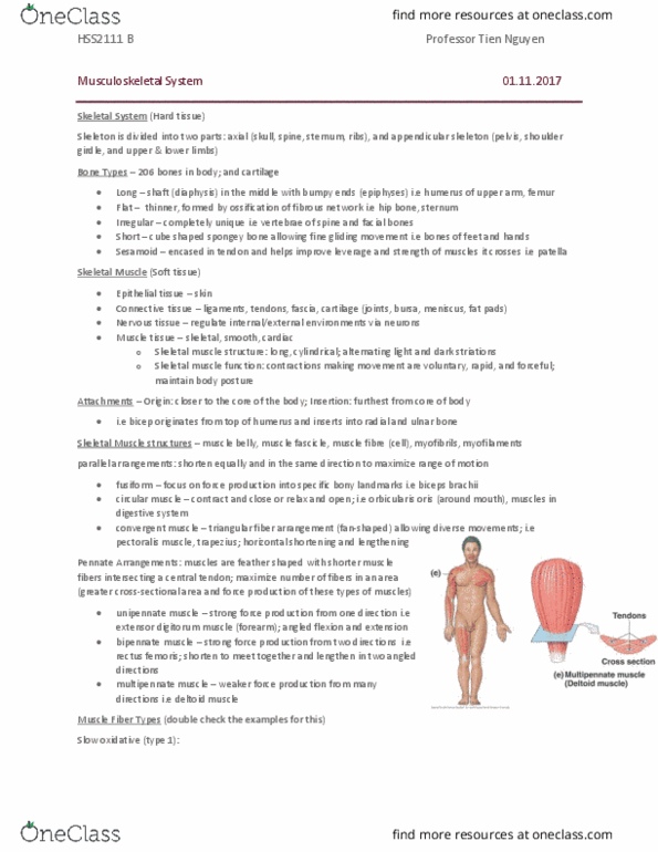
Skeleton is divided into two parts: axial (skull, spine, sternum, ribs), and appendicular skeleton (pelvis, shoulder girdle, and upper & lower limbs) Bone types 206 bones in body; and cartilage: long shaft in the middle with bumpy ends; i. e humerus of upper arm, femur thigh bone, flat i. e hip bone. Irregular i. e spine: short i. e foot, sesamoid i. e patella. Connective tissue ligaments, tendons, fascia, cartilage (joints, bursa, meniscus, fat pads) Nervous tissue regulate internal/external environments via neurons. Skeletal muscle structure: long, cylindrical; alternating light and dark striations. Skeletal muscle function: contractions making movement are voluntary, rapid, and forceful; maintain body posture. Attachments origin: closer to the core of the body; insertion: furthest from core of body i. e bicep originates from top of humerus and inserts into radial and ulnar bone. Multipennate muscle i. e deltoid muscle; multidirectional range of movement circular muscle orbicularis oris (around mouth); circular opening and closing. Muscle fiber types (double check the examples for this)