ANP 1106 Lecture Notes - Lecture 1: Extensor Digitorum Longus Muscle, Rectus Femoris Muscle, Biceps Femoris Muscle

15 Nov 2016
School
Department
Course
Professor

Anatomy 1106 Lecture 1: Chapter 9 (pg. 278-286)
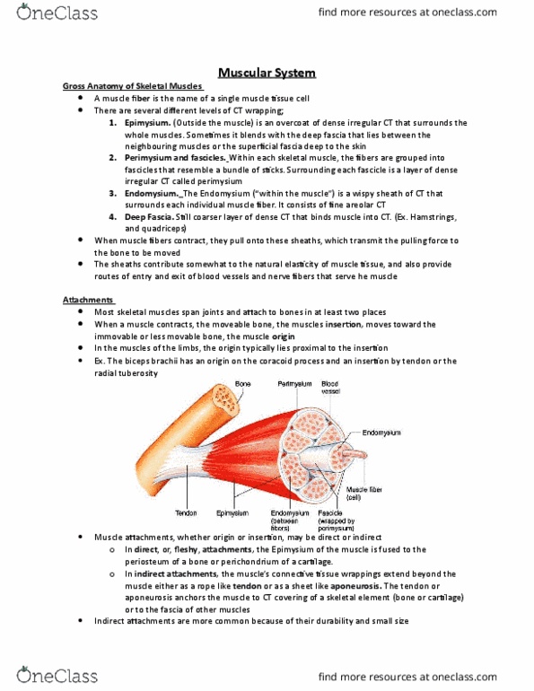
Layers of Connective Tissue;
1. Epimysium= the outermost layer, made up of dense irregular CT.
2. Perimysium and Fascicles= muscle fibers are grouped into fascicles (like
stick-bundles). Surrounding each fascicle is the perimysium (dense irregular
CT).
3. Endomysium= sheath of CT that surrounds each individual muscle fiber.
Consists of areolar CT.
Deep Fascia: coarser layer of dense CT that binds muscles into functional groups;
ex: hamstrings vs. quadriceps.
Attatchments
• Most skeletal muscles span joints and attach to bones in at least 2 places
• When a muscle contracts the muscle’s insertion moves toward the less
movable bone, the origin.
• Origins or insertions may be direct or indirect.
• Indirect are more common; they are like tendons, which anchors the muscle
to the CT covering of a skeletal element (like a bone or cartilage). They are
durable and small.
Arrangement of Fascicles
• All skeletal muscles have fascicles, but they vary in arrangements, causing
the shapes and functions of muscles to be different.
1. Circular: the fascicles are arranged in concentric rings. Muscles with this
arrangement surround external body opening, that closes by contracting, like
sphincters. Ex: muscles surrounding the eyes and mouth!
2. Convergent: muscle has a broad origin, and its fascicles converge toward a
single tendon of insertion. It’s triangular or fan shaped. Ex: the pectoralis
major.
3. Parallel: the length of the fascicles runs parallel to the long axis of the
muscle. They are strap-like (Sartorius muscle) OR spindle-shaped with an
expanded midsection (Biceps).
4. Pennate: The fascicles are short and they attach obliquely to a central
tendon that runs the length of the muscle. There are 3 forms:
a. Unipennate: the fascicles insert into only ONE side of the tendon
(extensor digitorum longus).
b. Bipennate: the fascicles insert into the tendon from opposite sides so
the muscle looks like a feather (rectus femoris).
c. Multipennate: looks like many feathers side by side, with all the
quills inserted into one tendon. Like the deltoid muscle.
Muscles in 4 functional groups:
find more resources at oneclass.com
find more resources at oneclass.com

1) Prime mover/ Agonist: is the muscle that has the major responsibility of
producing a specific movement. Ex: the Pectoralis major is a prime mover of
arm flexion.
2) Antagonist: the muscle that opposes the movement. Providing some
resistance, to prevent overshooting. Ex: flexion of the arm is antagonized by
the latissimus dorsi.
3) Synergist: most movements involve the action of one or more of these. They
help prime movers by adding a little extra force to the movement.
4) Fixators: When synergists immobilize a bone, or a muscles origin so that the
prime mover has a stable base to act on, they are called fixators! Ex: muscles
involves in posture!
How are the skeletal muscles named?
1) Location: Ex= the temporalis muscle is over the temporal bones
2) Shape: Ex= the trapezius muscles look like trapezoids
3) Size: Ex= Maximus is for largest, and minimus is for smallest
4) Direction of fibers: Ex= rectus (straight) muscles have fibers that run
parallel to the axis.
5) # Of origins: ex: the biceps has two heads/ origins
6) Location of the attachments: the origin is always named first! Ex:
sternocleidomastoid; has an origin on the sternum and clavicle, and
inserts on the mastoid process of the temporal bone.
7) Action: ex: flexor, extensor, and adductor.
Muscles of the head- Facial Expression:
• Epicranius: the main muscle of the scalp! Has:
a) Frontalis: which raises the eyebrows and wrinkles forehead
b) Occipitalis: pulls the scalp posteriorly
• Orbicularis oculi: it surrounds the rim of orbit; protects eyes from light and
injury- deals with blinking, squinting, and brings the eyebrows down
• Zygomaticus: smiling muscle
• Orbicularis oris: lips; multi-layered, closes, purses, and protrudes lips
• Mentalis: V-shaped pair- protrudes lower lip, wrinkles chin
• Buccinators: deep to masseter- whistling, sucking holds food in place when
chewing; especially in nursing infants.
• Platysma: helps depress mandible; tenses the skin of the neck
Muscles that move the tongue:
1) Genioglossus: Protracts the tongue; prevents the tongue from falling back.
2) Hyoglossus: depresses tongue, draws its sides inferiorly
3) Styloglossus: retracts and elevates the tongue
Muscles of Mastication:
1) Masseter: is the prime mover of the jaw, elevates the mandible
2) Temporalis: closes the jaw, elevates and retracts the mandible
find more resources at oneclass.com
find more resources at oneclass.com
Document Summary
Layers of connective tissue: epimysium= the outermost layer, made up of dense irregular ct, perimysium and fascicles= muscle fibers are grouped into fascicles (like stick-bundles). Surrounding each fascicle is the perimysium (dense irregular. Ct): endomysium= sheath of ct that surrounds each individual muscle fiber. Deep fascia: coarser layer of dense ct that binds muscles into functional groups; ex: hamstrings vs. quadriceps. Attatchments: most skeletal muscles span joints and attach to bones in at least 2 places, when a muscle contracts the muscle"s insertion moves toward the less movable bone, the origin, origins or insertions may be direct or indirect. Indirect are more common; they are like tendons, which anchors the muscle to the ct covering of a skeletal element (like a bone or cartilage). Arrangement of fascicles: all skeletal muscles have fascicles, but they vary in arrangements, causing the shapes and functions of muscles to be different, circular: the fascicles are arranged in concentric rings.