ANP 1105 Lecture Notes - Lecture 4: Warning Sign, Motor Unit, Heart Sounds

2 Nov 2017
School
Department
Course
Professor
Document Summary
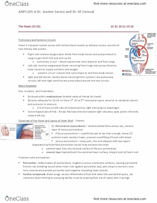
Extends obliquely for 12-14 cm from 2nd rib to 5th intercostal space. 2/3 of the mass is on the left side; right side lying on diaphragm. Broad flat base directed towards right shoulder, apex points towards left hip. 2 circuits: pulmonary circuit (right ventricle): short, low-pressure circulation, systemic circuit (left ventricle): long pathway with 5x resistance. 3 layers: pericardium: outer layer; sturdy, thick, strong, myocardium: middle layer; the actual muscle, endocardium: inner layer; single layer of endothelial cells. Cardiac muscle forms the bulk of the heart. Layer of endothelium + ct layer on inner myocardial surface. Continuous with endothelium of vessels creates a continuous lining. 2 exterior grooves: coronary sulcus (atrioventricular groove) and anterior- posterior interventricular sulcus. Pectinate muscles: bundles of muscles that project into the lumen of the atria (especially into the right atrium) Fossa ovalis: small depression that marks where the formen ovale existed in the fetal heart.