CAPS 391 Lecture Notes - Lecture 5: Femoral Vein, Posterior Interventricular Artery, Thymus

2 Feb 2019
School
Course
Professor
Document Summary
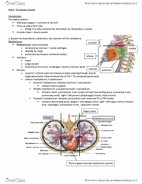
Divided into: superior, located between 1st rib and tiv/tv vertebrae. Inferior: anterior, located between sternum and pericardium, contains thymus gland, middle, contains heart, surrounding membranes is called pericardium, posterior, located between pericardium and bodies of vertebrae tv to txii, contains vagus nerves, esophagus and descending aorta. Fibrous (outer layer: thick layer of dense connective tissue, attaches the heart to the diaphragm via central tendon inferiorly and to the great vessels of the heart superiorly. Serous (inner layer: specialized in secretion of serous fluid, parietal, lines the inner surface of fibrous layer, visceral (epicardium, attaches to the surface of the heart. Pericardial cavity: tiny space between the parietal and visceral layers of the serous layer, filled with serous fluid that lubricates the heart surfaces to facilitate heart movements. Sa node: heart pacemaker, located on the right atrium wall, contracts the atrial walls. Av node: located on right side at the interatrial septum.