ACB 406.3 Lecture 16: Vision

6 Nov 2018
School
Department
Course
Professor
Document Summary
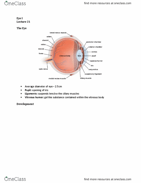
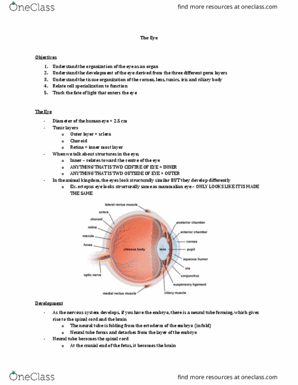
6 muscle suspend the eye in the bony eye socket (extraocular muscles of the eye) 3 layers: tunica fibrosa = corneoscleral coat (cornea and sclera, tunica vasculosa = uvea (choroid, ciliary body and iris, tunica intima = retina, and the epithelium of the ciliary body and iris. Corneal epithelium = non-cornified stratified squamous w/ microvilli (unusual: basal cells divide; cells linked by interdigitations and desmosomes, very rich in nerve endings. Basement membrane (bow(cid:373)a(cid:374)"s (cid:373)e(cid:373)(cid:271)ra(cid:374)e) bm of the epithelium: very thick bm made of collagen type iv. Corneal stroma = fibroblasts sandwiched b/w sheets of collagen and sulfated gags (glycosaminoglycans) arranged like plywood. Descemet"s (cid:373)e(cid:373)(cid:271)ra(cid:374)e = bm of the endothelium; is very thick. Corneal endothelium (not a true endothelium) = single layer of squamous cells: very active peripheral regeneration zone. *cornea is transparent because the layers of collagen are arranged at right angles of each other. Scelra parallel bundles of collagen oriented in various directions.