MBIO 1010 Lecture Notes - Lecture 3: Methylene Blue, Crystal Violet, Safranin

28 Dec 2017
School
Department
Course
Professor
Document Summary
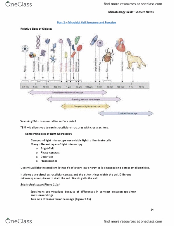
Chapter 2- microbial cell structure and function: microscopy, 2. 1: discovering cell structure: light microscopy, 2. 2: improving contrast in light microscopy, 2. 3 imaging cells in three dimensions, 2. 4 probing cell structure: electron microscopy. Relative sizes of objects: the size and wavelength of light is much much larger than the size of wavelength of electrons: electron microscopy is used to get super cool images. Improving contrast results in a better final image: staining improves contrast, dyes are organic compounds that bind to specific cellular materials, examples of common stains are methylene blue, safranin and crystal violet. Simple staining: on dye used to color specimen: chromophore coloured portion of a dye, bacteria have a negative charge: charge: cationic dyes attracted. Therefore the acid dyes are not used for samples that were heated before staining. 2: microscopy place drop of oil on slide; examine with 100x objective lens.