KIN 2320 Lecture Notes - Lecture 30: Carpal Tunnel, Ulnar Nerve, Lumbricals Of The Hand

28 Nov 2018
School
Department
Course
Professor

1
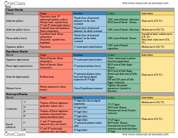
Short Muscles in Hand:
Lumbricals
1 + 2 – median nerve – flex metacarpophalangeal joints and extend interphalangeal joints 2nd – 5th fingers
- Unipennate – 1 direction
fibers
3 + 4 – deep branch of ulnar
nerve
- Bipennate - 2 feathers
Interossei - deep branch of
ulnar nerve
Dorsal – abduct 2-4th fingers
from axial line, act with
lumbricals to flex MCP joints
and extend IP joints
- Bipennate
Palmar – adduct 2, 4, and 5th
fingers and act with lumbricals
to flex MCP joints and extend IP
joints
- Unipennate
- Not on middle or thumb
Nerves of Hand:
Carpal tunnel: is the passageway deep to the flexor retinaculum between the tubercles of the scaphoid and
the trapezium bones on the lateral side and the pisiform and the hook of hamate on the medial side. Distal to
the carpal tunnel, the median nerve supplies two and half thenar muscles and the 1st and 2nd lumbricals.

2
The Ulnar Nerve
The ulnar nerve ends at the distal border of the flexor retinaculum by dividing into superficial and deep
branches. The superficial branch of the ulnar nerve supplies cutaneous branches to the anterior surfaces of
the medial one and a half fingers. The deep branch for the ulnar nerve supplies the hypothenar muscles, the
medial two lumbricals, the AD, the deep head of the FPB and all the interossei.
The deep branch also supplies several joints (wrist, intercarpal, carpometacarpal and intermetacarpal. The
ulnar nerve is referred to as the nerve of fine movements, because it innervates muscles that are concerned
with intricate hand movements.
Ulnar nerve – funny bone
The radial nerve – supplies no muscles in the hand.
Joints of Upper Limb:
Sternoclavicular joint: the SC joint is a synovial articulation between the sternal end of the clavicle and the
manubrium of the sternum and the 1st costal cartilage. The SC joint is a saddle type of joint but functions as a
ball and socket joint. Skeletal attachment of upper limb to body.
The SC joint is divided into two compartments by an articular disc.
Although the SC joint is extremely strong, it is significantly mobile to allow movements of the pectoral griddle
and upper limb. During full elevation of the limb, the clavicle, is raised to approximately a 60-degree angle.
The SC joint can also be moved anteriorly, posteriorly over a range of up to 25 to 30 degrees.
Document Summary
1 + 2 median nerve flex metacarpophalangeal joints and extend interphalangeal joints 2nd 5th fingers. 3 + 4 deep branch of ulnar nerve. Dorsal abduct 2-4th fingers from axial line, act with lumbricals to flex mcp joints and extend ip joints. Palmar adduct 2, 4, and 5th fingers and act with lumbricals to flex mcp joints and extend ip joints. Carpal tunnel: is the passageway deep to the flexor retinaculum between the tubercles of the scaphoid and the trapezium bones on the lateral side and the pisiform and the hook of hamate on the medial side. Distal to the carpal tunnel, the median nerve supplies two and half thenar muscles and the 1st and 2nd lumbricals. The ulnar nerve ends at the distal border of the flexor retinaculum by dividing into superficial and deep branches. The superficial branch of the ulnar nerve supplies cutaneous branches to the anterior surfaces of the medial one and a half fingers.