ZOO 3200 Lecture Notes - Lecture 6: Endoplasmic Reticulum, Rigor Mortis, Myocyte

2 May 2018
School
Department
Course
Professor

10/12/17
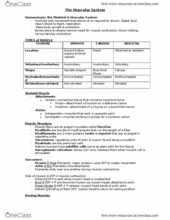
Categorization of muscle
!
Structural features of sarcomeres and myofilaments
○
Organization of skeletal muscles
!
The sliding-filament theory
!
The role of ATP in cross-bridge attachment
○
The role of Ca2+ in cross-bridge attachment
○
Cross-bridges and the production of force
!
Excitation-contraction coupling
!
Outline:
*see slide
○
Skeletal muscle (voluntary)
!
Cardiac muscle (involuntary)
!
Striated muscle:
○
Smooth muscle (involuntary)
!
Un-striated muscle:
○
3 muscle types can be classified in 2 different ways:
!
Muscles are either striated or un-striated (smooth) depending on whether or
not they have alternating dark and light bands
!
Sympathetic -fight or flight
!
Parasympathetic -basic physiological function
!
Enteric -movement of food through body
!
Autonomic branch:
○
*heart can be both sympathetic and parasympathetic
○
Muscles are either voluntary (motor division of efferent branch of PNS) or
involuntary (autonomic division of efferent branch) depending on the
division of the peripheral nervous system that innervates them
!
Categorization of Muscle
Muscles are anchored by tendons
○
Muscles are made up of multi-nucleated cells = muscle fibers
○
Muscle fibers are made of parallel subunits = myofibrils
○
Myofibrils consist of repeated units = sarcomeres
○
Each sarcomere has two types of myofilaments that are bound by Z
disks: actin thin filaments and myosin thick filaments
○
*see diagram on slide
○
Structural features of sarcomeres:
!
Thin filaments are composed of a two-strand actin helix, the
filamentous protein tropomyosin, and the troponin complex
○
The head region (cross-bridges) contain actin-and ATP-binding
sites
!
The thick filaments are composed of hundreds of identical myosin
proteins
○
Cross bridges extend from the thick filament and contact with the thin
filament during muscle contraction
○
Structural features of myofilaments:
!
Organization of Skeletal Muscles
*see diagram on slide
!
Shorter gaps between Z lines
○
Reduction in the H zone
○
Sarcomeres shorten during muscle contraction as thin filaments actively
slide along the thick filaments
!
Sliding-Filament Theory
Binding: hydrolysis of ATP causes myosin head to extend and attach to
actin
!
Power stroke: release of phosphate promotes myosin head rotation (pulls
actin)
!
*note mitochondria are required to supply ATP (in aerobic muscle)
○
Release: binding of ATP (from mitochondria) causes myosin to detach from
actin
!
*without ATP, myosin binds irreversibly to actin --> rigor mortis
!
Cross Bridges and the Production of Force: the role of ATP
When [Ca2+] is low, tropomyosin blocks the myosin-binding sites on action
!
Ca2+ enters cells when action potential reaches target cell
○
When [Ca2+] is high, Ca2+ binding to troponin (complex) removes an
attachment inhibition between myosin cross-bridges and thin filaments
!
Regulation of Muscle Contraction: the role of Ca2+
Tropomyosin complex blocks binding sites on actin site (TnI + TnT +
TnC)
○
This stop myosin head from connecting
○
Relaxed state:
!
Ca2+ binds to TnC causing TnI to move and reveal myosin binding sites on
actin
!
The Contractile Element
Muscle fibers contract when a postsynaptic end plate potential at the
neuromuscular junction causes a propagated action potential in the fiber
sarcolemma
!
Transverse (T) tubules conduct APs into the cell interior causing Ca2
+ release from the sarcoplasmic reticulum (intra-cellular calcium
store) that surround the myofibrils
○
How does an AP in the muscle fiber change the free [Ca2+] in the cytosol?
!
Voltage-sensitive DHPR and RyR work together, linking
depolarization of the T tubule to the opening of Ca2+ channels in the
sarcoplasmic reticulum membrane
○
Ca2+ in the sarcoplasmic reticulum membrane re-sequester Ca2+
from the cytosol
○
Calsequestrin inside the sarcoplasmic reticulum binds Ca2+ reducing
the free [Ca2+] inside the sarcoplasmic reticulum
○
Transporters and channels involved:
!
T-tubule lumen is positively charged
!
Myoplasm is negatively charged
!
Conditions at rest 1.
Ca2+ (from SR) binds to troponin on thin filament (ryanodine
receptor)
!
T-tubule lumen becomes negatively charged
!
Myoplasm becomes positively charged
!
Conditions when T-tubule depolarizes2.
Returns to resting potential, Ca2+ sequestered in SR by calcium
pump
!
Conditions when sarcolemma potential returns to resting value3.
Excitation-Contraction Coupling:
!
In cardiac muscles, entrance of extracellular Ca2+ through DHPR
triggers opening of RyR: Ca2+-induced Ca2+ release
○
*big difference between cardiac and skeletal muscle
○
In skeletal muscles, the DHPR and RyR physically interact: depolarization-
induced Ca2+ release
!
Excitation-Contraction Coupling
*see diagrams
!
Summary of Excitation-Co ntraction Couplin g
10/19/17
Role of the myofilament proteins in the evolution of muscle function
!
Twitches, summation and tetanus
!
Neuronal control of skeletal muscles
!
The force-velocity relationship
○
The length-tension relationship
○
Work of contraction
○
Mechanisms of muscle contraction
!
Energetics of muscle contraction
!
Outline:
*see figure
!
Higher the pCa --> lower the [Ca2+]
!
pCa50 = [Ca2+] required to generate half the maximum force
!
As temperature increases, heart becomes more sensitive to
calcium
!
Therefore, trout must have a higher sensitivity because they are
found in cold environments (are ectothermic)
!
Trout heart would remain in contracture at 37C
○
Can respond to calcium at lower temp
○
Trout heart is 10x more sensitive to Ca2+ as mammalian heart
!
All have similar pCa at their physiological temperature
○
Troat at 7C = frog at 22C = mammal = 37C
○
Red circle = physiologic temperature
!
Through vertebrate evolution, the heart has become less sensitive to Ca2+
See slide
!
Once myosin head binds, force is generated
!
The Contractile Element:
Logical place to look for mechanism of higher Ca2+ sensitivity
○
Ca2+ activated trigger --> change in protein shape
!
Only 13 sequence difference despite separation by ~300 million years
of evolution
○
Comparison of McTnC and salmonid cardiac troponin C (ScTnC)
!
cTnC cDNA in virus --> infect bacteria with virus
○
Grow large volumes of bacteria and force them to express protein
○
Purify protein from bacteria
○
Preform experiments on recombinant proteins
○
Methods: protein production
!
ScTnC is ~2.3 fold more sensitive to Ca2+ as McTnC
○
Sequential differences have functional consequences
○
ScTnC and McTnC at 21C
!
Manipulate McTnC cDNA in virus using site directed mutagenesis
○
4 residues: NIQD
!
Generates multiple mutant McTnC proteins containing different
residues from ScTnC
○
Methods: produce mutant McTnC proteins
!
NIQD McTnC increases the Ca2+ sensitivity of force generation in
mammalian cardiac myocytes
○
Ca2+ affinity of NIQD McTnC is similar to that of ScTnC
!
NIQD only in cTnC from ectotherms
○
Sequence changed with the evolution of endothermy to allow cardiac
function at warm temperatures
○
Comparisons of cTnCs from fish and mammalian species:
!
Shape of ScTnC makes it more readily activated
!
Sequence difference effect shape of molecules
○
Better able to work at low temperatures
!
ScTnC more flexible, easier to change conformation upon Ca2+
activation
○
Changes in sequence of McTnC impacted protein function, making it
better able to function at high temperatures (37C)
○
How are the identified residues increasing Ca2+ affinity:
!
Mammalian cTnC (McTnC)
What factors determine how much tension is produced by a twitch?
○
Relative amount of tension produced by a single AP = twitch
!
Why cant a single twitch elicit the maximum tension that a muscle fiber is
capable of?
!
The addition of tension due to repeated rapid stimulation = temporal
summation
!
Low frequency AP --> twitch
!
Maximum stimulation to muscles
○
Much faster
○
Only possible in extreme states
○
High frequency AP --> tetanus
!
Muscle is not allowed to reflax
○
*increased Ca2+ --> increasing tension
!
Twitches, Summation and Tetanus
Motor nerves contain 100-1000 motor neurons
!
A motor neuron branches to multiple muscle fibers, forming a motor unit
!
Each muscle fiber is innervated by only one motor neuron
!
Increasing AP frequency
○
Recruiting motor units
○
Recruiting fibers that have higher intensity of contraction
○
Neurons increase muscle tension by:
!
Neuronal Control of Skeletal Muscles
Max velocity when load =0 0
○
Elastic elements are stretched but muscle stays the same length
!
Isometric contraction when velocity = 0
○
As load increases the shortening velocity decreases
!
How can a sarcomere generate different amounts of force?
!
Why is there an inverse relationship between force generation and
contraction velocity?
!
Mechanisms of Muscle Contraction: the force-velocity relationship
How much tension a muscle can produce during contraction is related to its
resting length
!
*see slide
○
There is an optimal overlap of thick and thin filaments that produces the
maximum amount of tension during muscle contraction
!
The length-tension relationship for a sarcomere is strong evidence
supporting the sliding-filament theory
!
Increase number of cross bridges --> increase in force
!
Mechanics of Muscle Contraction: the length-tension relationship
*see slide
!
Work = force X distance
!
Muscle is able to shorten anymore with sufficient weight (W=0)
○
As you add weight, force of load increases
!
Greater cross section --> greater ability to do work
!
Muscle cell volume --> increase in mitochondria ?
○
The amount of work a muscle can do also depends on its volume
!
Mechanics of Muscle Contraction: work of contraction
ATP is needed for contraction1.
ATP is needed for relaxation2.
Creatine phosphate (very quick muscle movement)
○
Oxidative phosphorylation (long distance running)
○
Glycolysis (short distance running)
○
Three metabolic pathways supply the ATP:3.
Energetics of Muscle Contraction:
Characteristics of the three principle mechanisms of ATP production in vertebrate
muscle:
Creatine
Phosphate
Oxidative
Phosphorylation
Glycolysis
ATP synthesis
rate
Very fast Slow Fast
Yield of ATP Very low Very high Low
Primary Fuel
Use
Muscle CP Blood glucose &
FA
Muscle glycogen
Limitations Short duration Requires O2 and
slow
Low efficiency and
lactate acidosis
10/24/17
Energetics of muscle contraction
!
Muscle fatigue, recovery, and oxygen debt
!
Fiber types in vertebrate skeletal muscles
!
Skeletal muscle phenotypes and muscle performance
!
Muscle hypertrophy
○
Muscle atrophy
○
Skeletal muscle plasticity
!
Outline:
Hypothesis: creatine phosphate serves as a principal source of ATP during
the first seconds of burst exercise
!
Prediction: lowering the levels of creatine (CK) should interfere with burst
exercise
!
+/+ full CK activity
○
+/-heterozygous
○
-/- no CK activity
○
I/I 3-fold reduction in CK
○
I/- 6-fold reduction in CK
○
Methods: generation of mice with different CK enzyme activity
!
Result: ability of muscle to perform burst activity closely correlates with
CK activity
!
Genetic Engineering and the Physiological Role of Creatine Kinase
Fatigue has multiple causes depending on the type and duration of exercise
!
High-intensity short-term activity produces lactic acid which is an indicator
of fatigue
!
Fatigue associated with sustained exercise is partly due to inadequate
muscle glucose
!
Depletion of energy reserves
○
Ion disturbances
○
pH imbalance
○
In general, muscle fatigue results from:
!
Replenishing energy stores (using the Cori Cycle)
○
Re-establishing ion gradients (Ca2+ stores and pH)
○
Recovery involves:
!
*see slide
!
Muscle Fatigue and Recovery
The start of exercise is associated with an O2 deficit because demand is
larger than supply
!
Energy for recovery metabolism is provided by aerobic metabolism
!
= oxygen debt
○
Rates of O2 consumption remain elevated long after exercise has ceased
!
*see slide
!
Post-exercise Oxygen Recovery
Slow
Oxidative
Fast Oxidative
Glycolytic
Fast Glycolytic
Myosin ATPase
activity
Slow Fast Fast
Speed to reach peak
tension
Slow Intermediate-Fast Fast
Duration of Twitches Long Short Short
Rate of Ca2+ uptake
by SR
Slow-
Intermediate
High High
Resistance to fatigue High Intermediate Low
Number of
mitochondria
Many Many Few
Myoglobin content High High Low
Color Red Red White
Diameter of fiber Small Intermediate Large
Number of
surrounding
capillaries
Many Many Few
Levels of glycolytic
enzymes
Low Intermediate High
Ability to produce
ATP using oxidative
phosphorylation
High High Low
Force developed per
cross-sectional area
Low Intermediate High
Function in animal Posture Standing,
walking, rapid
repetitive
movements
Jumping, bursts
of high speed
locomotion
Frequency use by
animal
High Intermediate -
High
Low
As horse go from standing to trotting to galloping different fiber types are
used
!
More fast glycolytic fibers in swimmer (white)
○
More slow (and fast) oxidative fibers in long-distance cyclist
○
Comparing thigh muscle fiber composition to 50m sprint swimmer to long
distance cyclist:
!
Fiber Types in Vertebrate Skeletal Muscles
Most tetrapods are mosaics of different fiber types
!
Slow oxidative muscle fibers play a dominant role in endurance exercise
!
Slow
Oxidative
Fast Oxidative
Glycolytic
Fast
Glycolytic
Human Terms Type I Type IIa Type IIx
Myosin heavy-
chain isoform
Slow cross-
bridge cycling
Rapid cross-
bridge cycling
Rapid cross-
bridge cycling
Sarcoplasmic
Reticulum Ca2+-
ATPase
Slow Ca2+
uptake
Fast Ca2+
uptake
Fast Ca2+
uptake
Speed of
Contraction
Slow Fast Fast
Fast muscle fibers play a dominant role in strength or resistance exercise
!
Skeletal Muscle Phenotypes and Muscle Performance
Increased proportion of type IIa, decreased proportion of type IIx
!
Endurance training increased capillary density via angiogenesis (increase
amount of blood flow --> O2 and other molecules)
!
Aggregates of subsarcolemmal mitochondria appear more frequently
after training
○
Intercellular lipid inclusions are seen before and after endurance
training
○
Capillaries, subsarcolemma, and interfibrillar mitochondria are more
numerous after training
!
Endurance training increased number and size of mitochondria
!
Endurance training increased the volume of muscle fiber occupied by lipid
droplets
!
Effects of Endurance Training on Muscle Composition
Resistance training increased proportion of type IIa, decreased porportion of
type IIx
!
Resistance training increased the diameter of individual fibers by
hypertrophy
!
Decreases type I and II
○
Post-detraining conditions are optimal for sprinters
!
Effects of Resistance Training on Muscle Composition
10/26/17
Sound production
○
Flying
○
Heat production in fish
○
Adaptations of muscles for diverse activities
!
Outline:
Readings: insect flight, & "the quest for speed"
*see slide
!
Short Ca2+ transients
!
Quick cross-bridge cycle activity
!
Two conditions must be met:
!
Sonic muscles around the swim bladder of toadfish undergo contraction-
relaxation cycles at 200-300Hz without going into tetanus
!
Adaptations of Muscles for Sound Production
% myofibrillar
!
% mitochondrial
!
% sarcoplasmic reticulum
!
Skeletal muscles are a composite of three components (that determine
muscle fiber volume)
!
The only way one function can be increased is that the expense of
another
!
Flight requires the production of continuous (aerobic) high power output at
high contraction frequencies
!
Increasing muscle temperature*
!
Skeletal adaptations*
!
Whole body VO2 increases
!
Similar VO2 per um^2 of mitochondria
!
Mitochondrial adaptations
!
Adaptations:
!
These animals cannot fly at frequencies greater than 100Hz
!
The flight muscles of vertebrates and some insects are synchronous
!
This breakthrough design enables frequencies up to 1000Hz due
to space and energy-saving solutions
!
The flight muscles of most insects are asynchronous
!
*see slide(s)
!
In asynchronous flight muscles, due to reduced Ca2+ handling,
less space is occupied by the sarcoplasmic reticulum and
mitochondria, and so more space is occupied by myofibrils
!
A single action potential in asynchronous flight muscle initiates a
series of contractions that are triggered by stretch; the frequency of
contractions is not synchronized with the frequency of action
potentials
!
Insect Flight:
!
Adaptation of Muscles for Flying
Ex. Blue Marlin and Butterfly mackerel
!
The brain of a billfish is 10-15C warmer than the environment
!
Brain and eye are warmed by a heater organ in billfish and mackerel
!
Composed of thousands of closely intermingled veins and arteries
functioning as a countercurrent heat exchanger
!
Allows for cold O2 rich blood from gills to come in close contact with
warm O2 depleted blood from the tissue
!
Blood from the gill is warmed and heat loss from the tissue is
minimized
!
Rete Mirabile -specialized structure in the circulatory system
!
*see 40x magnification of cross section of caecum rete from bluefin tuna
!
*see following slide
!
Billfish -modified superior rectus (eye muscle)
!
Butterfly mackerel -lateral rectus
!
Muscle cells have been modified to produce heat without contracting
!
Artery provides dedicated source of blood
!
The heater organ is a modified eye muscle
!
Contains mitochondria, contractile apparatus, and sarcoplasmic
reticulum
!
Increases ability to produce ATP
"
Increase in mitochondria content (60% of cell volume)
!
Increases ability to store and release Ca2+
"
Increase in SR content
!
Increases ability to release Ca2+ into cell
"
Proliferation of T-tubules
!
Loss of contractive apparatus:
!
Futile Ca2+ cycling: moving Ca2+ to burn ATP --> heat produced as
a by-product
!
Heater cell is modified skeletal muscle cell
!
As the water temperature fluctuates greatly throughout the day, the sword
fish maintains its cranial temperature within 5C
!
Cranial Endothermy
*=general adaptations to
flying
Muscle Physiology
#$%&'()*+, -./012&, 34+,4536
34789,:;

10/12/17
Categorization of muscle
!
Structural features of sarcomeres and myofilaments
○
Organization of skeletal muscles
!
The sliding-filament theory
!
The role of ATP in cross-bridge attachment
○
The role of Ca2+ in cross-bridge attachment
○
Cross-bridges and the production of force
!
Excitation-contraction coupling
!
Outline:
*see slide
○
Skeletal muscle (voluntary)
!
Cardiac muscle (involuntary)
!
Striated muscle:
○
Smooth muscle (involuntary)
!
Un-striated muscle:
○
3 muscle types can be classified in 2 different ways:
!
Muscles are either striated or un-striated (smooth) depending on whether or
not they have alternating dark and light bands
!
Sympathetic -fight or flight
!
Parasympathetic -basic physiological function
!
Enteric -movement of food through body
!
Autonomic branch:
○
*heart can be both sympathetic and parasympathetic
○
Muscles are either voluntary (motor division of efferent branch of PNS) or
involuntary (autonomic division of efferent branch) depending on the
division of the peripheral nervous system that innervates them
!
Categorization of Muscle
Muscles are anchored by tendons
○
Muscles are made up of multi-nucleated cells = muscle fibers
○
Muscle fibers are made of parallel subunits = myofibrils
○
Myofibrils consist of repeated units = sarcomeres
○
Each sarcomere has two types of myofilaments that are bound by Z
disks: actin thin filaments and myosin thick filaments
○
*see diagram on slide
○
Structural features of sarcomeres:
!
Thin filaments are composed of a two-strand actin helix, the
filamentous protein tropomyosin, and the troponin complex
○
The head region (cross-bridges) contain actin-and ATP-binding
sites
!
The thick filaments are composed of hundreds of identical myosin
proteins
○
Cross bridges extend from the thick filament and contact with the thin
filament during muscle contraction
○
Structural features of myofilaments:
!
Organization of Skeletal Muscles
*see diagram on slide
!
Shorter gaps between Z lines
○
Reduction in the H zone
○
Sarcomeres shorten during muscle contraction as thin filaments actively
slide along the thick filaments
!
Sliding-Filament Theory
Binding: hydrolysis of ATP causes myosin head to extend and attach to
actin
!
Power stroke: release of phosphate promotes myosin head rotation (pulls
actin)
!
*note mitochondria are required to supply ATP (in aerobic muscle)
○
Release: binding of ATP (from mitochondria) causes myosin to detach from
actin
!
*without ATP, myosin binds irreversibly to actin --> rigor mortis
!
Cross Bridges and the Production of Force: the role of ATP
When [Ca2+] is low, tropomyosin blocks the myosin-binding sites on action
!
Ca2+ enters cells when action potential reaches target cell
○
When [Ca2+] is high, Ca2+ binding to troponin (complex) removes an
attachment inhibition between myosin cross-bridges and thin filaments
!
Regulation of Muscle Contraction: the role of Ca2+
Tropomyosin complex blocks binding sites on actin site (TnI + TnT +
TnC)
○
This stop myosin head from connecting
○
Relaxed state:
!
Ca2+ binds to TnC causing TnI to move and reveal myosin binding sites on
actin
!
The Contractile Element
Muscle fibers contract when a postsynaptic end plate potential at the
neuromuscular junction causes a propagated action potential in the fiber
sarcolemma
!
Transverse (T) tubules conduct APs into the cell interior causing Ca2
+ release from the sarcoplasmic reticulum (intra-cellular calcium
store) that surround the myofibrils
○
How does an AP in the muscle fiber change the free [Ca2+] in the cytosol?
!
Voltage-sensitive DHPR and RyR work together, linking
depolarization of the T tubule to the opening of Ca2+ channels in the
sarcoplasmic reticulum membrane
○
Ca2+ in the sarcoplasmic reticulum membrane re-sequester Ca2+
from the cytosol
○
Calsequestrin inside the sarcoplasmic reticulum binds Ca2+ reducing
the free [Ca2+] inside the sarcoplasmic reticulum
○
Transporters and channels involved:
!
T-tubule lumen is positively charged
!
Myoplasm is negatively charged
!
Conditions at rest 1.
Ca2+ (from SR) binds to troponin on thin filament (ryanodine
receptor)
!
T-tubule lumen becomes negatively charged
!
Myoplasm becomes positively charged
!
Conditions when T-tubule depolarizes2.
Returns to resting potential, Ca2+ sequestered in SR by calcium
pump
!
Conditions when sarcolemma potential returns to resting value3.
Excitation-Contraction Coupling:
!
In cardiac muscles, entrance of extracellular Ca2+ through DHPR
triggers opening of RyR: Ca2+-induced Ca2+ release
○
*big difference between cardiac and skeletal muscle
○
In skeletal muscles, the DHPR and RyR physically interact: depolarization-
induced Ca2+ release
!
Excitation-Contraction Coupling
*see diagrams
!
Summary of Excitation-Co ntraction Couplin g
10/19/17
Role of the myofilament proteins in the evolution of muscle function
!
Twitches, summation and tetanus
!
Neuronal control of skeletal muscles
!
The force-velocity relationship
○
The length-tension relationship
○
Work of contraction
○
Mechanisms of muscle contraction
!
Energetics of muscle contraction
!
Outline:
*see figure
!
Higher the pCa --> lower the [Ca2+]
!
pCa50 = [Ca2+] required to generate half the maximum force
!
As temperature increases, heart becomes more sensitive to
calcium
!
Therefore, trout must have a higher sensitivity because they are
found in cold environments (are ectothermic)
!
Trout heart would remain in contracture at 37C
○
Can respond to calcium at lower temp
○
Trout heart is 10x more sensitive to Ca2+ as mammalian heart
!
All have similar pCa at their physiological temperature
○
Troat at 7C = frog at 22C = mammal = 37C
○
Red circle = physiologic temperature
!
Through vertebrate evolution, the heart has become less sensitive to Ca2+
See slide
!
Once myosin head binds, force is generated
!
The Contractile Element:
Logical place to look for mechanism of higher Ca2+ sensitivity
○
Ca2+ activated trigger --> change in protein shape
!
Only 13 sequence difference despite separation by ~300 million years
of evolution
○
Comparison of McTnC and salmonid cardiac troponin C (ScTnC)
!
cTnC cDNA in virus --> infect bacteria with virus
○
Grow large volumes of bacteria and force them to express protein
○
Purify protein from bacteria
○
Preform experiments on recombinant proteins
○
Methods: protein production
!
ScTnC is ~2.3 fold more sensitive to Ca2+ as McTnC
○
Sequential differences have functional consequences
○
ScTnC and McTnC at 21C
!
Manipulate McTnC cDNA in virus using site directed mutagenesis
○
4 residues: NIQD
!
Generates multiple mutant McTnC proteins containing different
residues from ScTnC
○
Methods: produce mutant McTnC proteins
!
NIQD McTnC increases the Ca2+ sensitivity of force generation in
mammalian cardiac myocytes
○
Ca2+ affinity of NIQD McTnC is similar to that of ScTnC
!
NIQD only in cTnC from ectotherms
○
Sequence changed with the evolution of endothermy to allow cardiac
function at warm temperatures
○
Comparisons of cTnCs from fish and mammalian species:
!
Shape of ScTnC makes it more readily activated
!
Sequence difference effect shape of molecules
○
Better able to work at low temperatures
!
ScTnC more flexible, easier to change conformation upon Ca2+
activation
○
Changes in sequence of McTnC impacted protein function, making it
better able to function at high temperatures (37C)
○
How are the identified residues increasing Ca2+ affinity:
!
Mammalian cTnC (McTnC)
What factors determine how much tension is produced by a twitch?
○
Relative amount of tension produced by a single AP = twitch
!
Why cant a single twitch elicit the maximum tension that a muscle fiber is
capable of?
!
The addition of tension due to repeated rapid stimulation = temporal
summation
!
Low frequency AP --> twitch
!
Maximum stimulation to muscles
○
Much faster
○
Only possible in extreme states
○
High frequency AP --> tetanus
!
Muscle is not allowed to reflax
○
*increased Ca2+ --> increasing tension
!
Twitches, Summation and Tetanus
Motor nerves contain 100-1000 motor neurons
!
A motor neuron branches to multiple muscle fibers, forming a motor unit
!
Each muscle fiber is innervated by only one motor neuron
!
Increasing AP frequency
○
Recruiting motor units
○
Recruiting fibers that have higher intensity of contraction
○
Neurons increase muscle tension by:
!
Neuronal Control of Skeletal Muscles
Max velocity when load =0 0
○
Elastic elements are stretched but muscle stays the same length
!
Isometric contraction when velocity = 0
○
As load increases the shortening velocity decreases
!
How can a sarcomere generate different amounts of force?
!
Why is there an inverse relationship between force generation and
contraction velocity?
!
Mechanisms of Muscle Contraction: the force-velocity relationship
How much tension a muscle can produce during contraction is related to its
resting length
!
*see slide
○
There is an optimal overlap of thick and thin filaments that produces the
maximum amount of tension during muscle contraction
!
The length-tension relationship for a sarcomere is strong evidence
supporting the sliding-filament theory
!
Increase number of cross bridges --> increase in force
!
Mechanics of Muscle Contraction: the length-tension relationship
*see slide
!
Work = force X distance
!
Muscle is able to shorten anymore with sufficient weight (W=0)
○
As you add weight, force of load increases
!
Greater cross section --> greater ability to do work
!
Muscle cell volume --> increase in mitochondria ?
○
The amount of work a muscle can do also depends on its volume
!
Mechanics of Muscle Contraction: work of contraction
ATP is needed for contraction1.
ATP is needed for relaxation2.
Creatine phosphate (very quick muscle movement)
○
Oxidative phosphorylation (long distance running)
○
Glycolysis (short distance running)
○
Three metabolic pathways supply the ATP:3.
Energetics of Muscle Contraction:
Characteristics of the three principle mechanisms of ATP production in vertebrate
muscle:
Creatine
Phosphate
Oxidative
Phosphorylation
Glycolysis
ATP synthesis
rate
Very fast Slow Fast
Yield of ATP Very low Very high Low
Primary Fuel
Use
Muscle CP Blood glucose &
FA
Muscle glycogen
Limitations Short duration Requires O2 and
slow
Low efficiency and
lactate acidosis
10/24/17
Energetics of muscle contraction
!
Muscle fatigue, recovery, and oxygen debt
!
Fiber types in vertebrate skeletal muscles
!
Skeletal muscle phenotypes and muscle performance
!
Muscle hypertrophy
○
Muscle atrophy
○
Skeletal muscle plasticity
!
Outline:
Hypothesis: creatine phosphate serves as a principal source of ATP during
the first seconds of burst exercise
!
Prediction: lowering the levels of creatine (CK) should interfere with burst
exercise
!
+/+ full CK activity
○
+/-heterozygous
○
-/- no CK activity
○
I/I 3-fold reduction in CK
○
I/- 6-fold reduction in CK
○
Methods: generation of mice with different CK enzyme activity
!
Result: ability of muscle to perform burst activity closely correlates with
CK activity
!
Genetic Engineering and the Physiological Role of Creatine Kinase
Fatigue has multiple causes depending on the type and duration of exercise
!
High-intensity short-term activity produces lactic acid which is an indicator
of fatigue
!
Fatigue associated with sustained exercise is partly due to inadequate
muscle glucose
!
Depletion of energy reserves
○
Ion disturbances
○
pH imbalance
○
In general, muscle fatigue results from:
!
Replenishing energy stores (using the Cori Cycle)
○
Re-establishing ion gradients (Ca2+ stores and pH)
○
Recovery involves:
!
*see slide
!
Muscle Fatigue and Recovery
The start of exercise is associated with an O2 deficit because demand is
larger than supply
!
Energy for recovery metabolism is provided by aerobic metabolism
!
= oxygen debt
○
Rates of O2 consumption remain elevated long after exercise has ceased
!
*see slide
!
Post-exercise Oxygen Recovery
Slow
Oxidative
Fast Oxidative
Glycolytic
Fast Glycolytic
Myosin ATPase
activity
Slow Fast Fast
Speed to reach peak
tension
Slow Intermediate-Fast Fast
Duration of Twitches Long Short Short
Rate of Ca2+ uptake
by SR
Slow-
Intermediate
High High
Resistance to fatigue High Intermediate Low
Number of
mitochondria
Many Many Few
Myoglobin content High High Low
Color Red Red White
Diameter of fiber Small Intermediate Large
Number of
surrounding
capillaries
Many Many Few
Levels of glycolytic
enzymes
Low Intermediate High
Ability to produce
ATP using oxidative
phosphorylation
High High Low
Force developed per
cross-sectional area
Low Intermediate High
Function in animal Posture Standing,
walking, rapid
repetitive
movements
Jumping, bursts
of high speed
locomotion
Frequency use by
animal
High Intermediate -
High
Low
As horse go from standing to trotting to galloping different fiber types are
used
!
More fast glycolytic fibers in swimmer (white)
○
More slow (and fast) oxidative fibers in long-distance cyclist
○
Comparing thigh muscle fiber composition to 50m sprint swimmer to long
distance cyclist:
!
Fiber Types in Vertebrate Skeletal Muscles
Most tetrapods are mosaics of different fiber types
!
Slow oxidative muscle fibers play a dominant role in endurance exercise
!
Slow
Oxidative
Fast Oxidative
Glycolytic
Fast
Glycolytic
Human Terms Type I Type IIa Type IIx
Myosin heavy-
chain isoform
Slow cross-
bridge cycling
Rapid cross-
bridge cycling
Rapid cross-
bridge cycling
Sarcoplasmic
Reticulum Ca2+-
ATPase
Slow Ca2+
uptake
Fast Ca2+
uptake
Fast Ca2+
uptake
Speed of
Contraction
Slow Fast Fast
Fast muscle fibers play a dominant role in strength or resistance exercise
!
Skeletal Muscle Phenotypes and Muscle Performance
Increased proportion of type IIa, decreased proportion of type IIx
!
Endurance training increased capillary density via angiogenesis (increase
amount of blood flow --> O2 and other molecules)
!
Aggregates of subsarcolemmal mitochondria appear more frequently
after training
○
Intercellular lipid inclusions are seen before and after endurance
training
○
Capillaries, subsarcolemma, and interfibrillar mitochondria are more
numerous after training
!
Endurance training increased number and size of mitochondria
!
Endurance training increased the volume of muscle fiber occupied by lipid
droplets
!
Effects of Endurance Training on Muscle Composition
Resistance training increased proportion of type IIa, decreased porportion of
type IIx
!
Resistance training increased the diameter of individual fibers by
hypertrophy
!
Decreases type I and II
○
Post-detraining conditions are optimal for sprinters
!
Effects of Resistance Training on Muscle Composition
10/26/17
Sound production
○
Flying
○
Heat production in fish
○
Adaptations of muscles for diverse activities
!
Outline:
Readings: insect flight, & "the quest for speed"
*see slide
!
Short Ca2+ transients
!
Quick cross-bridge cycle activity
!
Two conditions must be met:
!
Sonic muscles around the swim bladder of toadfish undergo contraction-
relaxation cycles at 200-300Hz without going into tetanus
!
Adaptations of Muscles for Sound Production
% myofibrillar
!
% mitochondrial
!
% sarcoplasmic reticulum
!
Skeletal muscles are a composite of three components (that determine
muscle fiber volume)
!
The only way one function can be increased is that the expense of
another
!
Flight requires the production of continuous (aerobic) high power output at
high contraction frequencies
!
Increasing muscle temperature*
!
Skeletal adaptations*
!
Whole body VO2 increases
!
Similar VO2 per um^2 of mitochondria
!
Mitochondrial adaptations
!
Adaptations:
!
These animals cannot fly at frequencies greater than 100Hz
!
The flight muscles of vertebrates and some insects are synchronous
!
This breakthrough design enables frequencies up to 1000Hz due
to space and energy-saving solutions
!
The flight muscles of most insects are asynchronous
!
*see slide(s)
!
In asynchronous flight muscles, due to reduced Ca2+ handling,
less space is occupied by the sarcoplasmic reticulum and
mitochondria, and so more space is occupied by myofibrils
!
A single action potential in asynchronous flight muscle initiates a
series of contractions that are triggered by stretch; the frequency of
contractions is not synchronized with the frequency of action
potentials
!
Insect Flight:
!
Adaptation of Muscles for Flying
Ex. Blue Marlin and Butterfly mackerel
!
The brain of a billfish is 10-15C warmer than the environment
!
Brain and eye are warmed by a heater organ in billfish and mackerel
!
Composed of thousands of closely intermingled veins and arteries
functioning as a countercurrent heat exchanger
!
Allows for cold O2 rich blood from gills to come in close contact with
warm O2 depleted blood from the tissue
!
Blood from the gill is warmed and heat loss from the tissue is
minimized
!
Rete Mirabile -specialized structure in the circulatory system
!
*see 40x magnification of cross section of caecum rete from bluefin tuna
!
*see following slide
!
Billfish -modified superior rectus (eye muscle)
!
Butterfly mackerel -lateral rectus
!
Muscle cells have been modified to produce heat without contracting
!
Artery provides dedicated source of blood
!
The heater organ is a modified eye muscle
!
Contains mitochondria, contractile apparatus, and sarcoplasmic
reticulum
!
Increases ability to produce ATP
"
Increase in mitochondria content (60% of cell volume)
!
Increases ability to store and release Ca2+
"
Increase in SR content
!
Increases ability to release Ca2+ into cell
"
Proliferation of T-tubules
!
Loss of contractive apparatus:
!
Futile Ca2+ cycling: moving Ca2+ to burn ATP --> heat produced as
a by-product
!
Heater cell is modified skeletal muscle cell
!
As the water temperature fluctuates greatly throughout the day, the sword
fish maintains its cranial temperature within 5C
!
Cranial Endothermy
*=general adaptations to
flying
Muscle Physiology
#$%&'()*+, -./012&, 34+,4536 34789,:;

10/12/17
Categorization of muscle
!
Structural features of sarcomeres and myofilaments
○
Organization of skeletal muscles
!
The sliding-filament theory
!
The role of ATP in cross-bridge attachment
○
The role of Ca2+ in cross-bridge attachment
○
Cross-bridges and the production of force
!
Excitation-contraction coupling
!
Outline:
*see slide
○
Skeletal muscle (voluntary)
!
Cardiac muscle (involuntary)
!
Striated muscle:
○
Smooth muscle (involuntary)
!
Un-striated muscle:
○
3 muscle types can be classified in 2 different ways:
!
Muscles are either striated or un-striated (smooth) depending on whether or
not they have alternating dark and light bands
!
Sympathetic -fight or flight
!
Parasympathetic -basic physiological function
!
Enteric -movement of food through body
!
Autonomic branch:
○
*heart can be both sympathetic and parasympathetic
○
Muscles are either voluntary (motor division of efferent branch of PNS) or
involuntary (autonomic division of efferent branch) depending on the
division of the peripheral nervous system that innervates them
!
Categorization of Muscle
Muscles are anchored by tendons
○
Muscles are made up of multi-nucleated cells = muscle fibers
○
Muscle fibers are made of parallel subunits = myofibrils
○
Myofibrils consist of repeated units = sarcomeres
○
Each sarcomere has two types of myofilaments that are bound by Z
disks: actin thin filaments and myosin thick filaments
○
*see diagram on slide
○
Structural features of sarcomeres:
!
Thin filaments are composed of a two-strand actin helix, the
filamentous protein tropomyosin, and the troponin complex
○
The head region (cross-bridges) contain actin-and ATP-binding
sites
!
The thick filaments are composed of hundreds of identical myosin
proteins
○
Cross bridges extend from the thick filament and contact with the thin
filament during muscle contraction
○
Structural features of myofilaments:
!
Organization of Skeletal Muscles
*see diagram on slide
!
Shorter gaps between Z lines
○
Reduction in the H zone
○
Sarcomeres shorten during muscle contraction as thin filaments actively
slide along the thick filaments
!
Sliding-Filament Theory
Binding: hydrolysis of ATP causes myosin head to extend and attach to
actin
!
Power stroke: release of phosphate promotes myosin head rotation (pulls
actin)
!
*note mitochondria are required to supply ATP (in aerobic muscle)
○
Release: binding of ATP (from mitochondria) causes myosin to detach from
actin
!
*without ATP, myosin binds irreversibly to actin --> rigor mortis
!
Cross Bridges and the Production of Force: the role of ATP
When [Ca2+] is low, tropomyosin blocks the myosin-binding sites on action
!
Ca2+ enters cells when action potential reaches target cell
○
When [Ca2+] is high, Ca2+ binding to troponin (complex) removes an
attachment inhibition between myosin cross-bridges and thin filaments
!
Regulation of Muscle Contraction: the role of Ca2+
Tropomyosin complex blocks binding sites on actin site (TnI + TnT +
TnC)
○
This stop myosin head from connecting
○
Relaxed state:
!
Ca2+ binds to TnC causing TnI to move and reveal myosin binding sites on
actin
!
The Contractile Element
Muscle fibers contract when a postsynaptic end plate potential at the
neuromuscular junction causes a propagated action potential in the fiber
sarcolemma
!
Transverse (T) tubules conduct APs into the cell interior causing Ca2
+ release from the sarcoplasmic reticulum (intra-cellular calcium
store) that surround the myofibrils
○
How does an AP in the muscle fiber change the free [Ca2+] in the cytosol?
!
Voltage-sensitive DHPR and RyR work together, linking
depolarization of the T tubule to the opening of Ca2+ channels in the
sarcoplasmic reticulum membrane
○
Ca2+ in the sarcoplasmic reticulum membrane re-sequester Ca2+
from the cytosol
○
Calsequestrin inside the sarcoplasmic reticulum binds Ca2+ reducing
the free [Ca2+] inside the sarcoplasmic reticulum
○
Transporters and channels involved:
!
T-tubule lumen is positively charged
!
Myoplasm is negatively charged
!
Conditions at rest
1.
Ca2+ (from SR) binds to troponin on thin filament (ryanodine
receptor)
!
T-tubule lumen becomes negatively charged
!
Myoplasm becomes positively charged
!
Conditions when T-tubule depolarizes
2.
Returns to resting potential, Ca2+ sequestered in SR by calcium
pump
!
Conditions when sarcolemma potential returns to resting value
3.
Excitation-Contraction Coupling:
!
In cardiac muscles, entrance of extracellular Ca2+ through DHPR
triggers opening of RyR: Ca2+-induced Ca2+ release
○
*big difference between cardiac and skeletal muscle
○
In skeletal muscles, the DHPR and RyR physically interact: depolarization-
induced Ca2+ release
!
Excitation-Contraction Coupling
*see diagrams
!
Summary of Excitation-Co ntraction Couplin g
10/19/17
Role of the myofilament proteins in the evolution of muscle function
!
Twitches, summation and tetanus
!
Neuronal control of skeletal muscles
!
The force-velocity relationship
○
The length-tension relationship
○
Work of contraction
○
Mechanisms of muscle contraction
!
Energetics of muscle contraction
!
Outline:
*see figure
!
Higher the pCa --> lower the [Ca2+]
!
pCa50 = [Ca2+] required to generate half the maximum force
!
As temperature increases, heart becomes more sensitive to
calcium
!
Therefore, trout must have a higher sensitivity because they are
found in cold environments (are ectothermic)
!
Trout heart would remain in contracture at 37C
○
Can respond to calcium at lower temp
○
Trout heart is 10x more sensitive to Ca2+ as mammalian heart
!
All have similar pCa at their physiological temperature
○
Troat at 7C = frog at 22C = mammal = 37C
○
Red circle = physiologic temperature
!
Through vertebrate evolution, the heart has become less sensitive to Ca2+
See slide
!
Once myosin head binds, force is generated
!
The Contractile Element:
Logical place to look for mechanism of higher Ca2+ sensitivity
○
Ca2+ activated trigger --> change in protein shape
!
Only 13 sequence difference despite separation by ~300 million years
of evolution
○
Comparison of McTnC and salmonid cardiac troponin C (ScTnC)
!
cTnC cDNA in virus --> infect bacteria with virus
○
Grow large volumes of bacteria and force them to express protein
○
Purify protein from bacteria
○
Preform experiments on recombinant proteins
○
Methods: protein production
!
ScTnC is ~2.3 fold more sensitive to Ca2+ as McTnC
○
Sequential differences have functional consequences
○
ScTnC and McTnC at 21C
!
Manipulate McTnC cDNA in virus using site directed mutagenesis
○
4 residues: NIQD
!
Generates multiple mutant McTnC proteins containing different
residues from ScTnC
○
Methods: produce mutant McTnC proteins
!
NIQD McTnC increases the Ca2+ sensitivity of force generation in
mammalian cardiac myocytes
○
Ca2+ affinity of NIQD McTnC is similar to that of ScTnC
!
NIQD only in cTnC from ectotherms
○
Sequence changed with the evolution of endothermy to allow cardiac
function at warm temperatures
○
Comparisons of cTnCs from fish and mammalian species:
!
Shape of ScTnC makes it more readily activated
!
Sequence difference effect shape of molecules
○
Better able to work at low temperatures
!
ScTnC more flexible, easier to change conformation upon Ca2+
activation
○
Changes in sequence of McTnC impacted protein function, making it
better able to function at high temperatures (37C)
○
How are the identified residues increasing Ca2+ affinity:
!
Mammalian cTnC (McTnC)
What factors determine how much tension is produced by a twitch?
○
Relative amount of tension produced by a single AP = twitch
!
Why cant a single twitch elicit the maximum tension that a muscle fiber is
capable of?
!
The addition of tension due to repeated rapid stimulation = temporal
summation
!
Low frequency AP --> twitch
!
Maximum stimulation to muscles
○
Much faster
○
Only possible in extreme states
○
High frequency AP --> tetanus
!
Muscle is not allowed to reflax
○
*increased Ca2+ --> increasing tension
!
Twitches, Summation and Tetanus
Motor nerves contain 100-1000 motor neurons
!
A motor neuron branches to multiple muscle fibers, forming a motor unit
!
Each muscle fiber is innervated by only one motor neuron
!
Increasing AP frequency
○
Recruiting motor units
○
Recruiting fibers that have higher intensity of contraction
○
Neurons increase muscle tension by:
!
Neuronal Control of Skeletal Muscles
Max velocity when load =0 0
○
Elastic elements are stretched but muscle stays the same length
!
Isometric contraction when velocity = 0
○
As load increases the shortening velocity decreases
!
How can a sarcomere generate different amounts of force?
!
Why is there an inverse relationship between force generation and
contraction velocity?
!
Mechanisms of Muscle Contraction: the force-velocity relationship
How much tension a muscle can produce during contraction is related to its
resting length
!
*see slide
○
There is an optimal overlap of thick and thin filaments that produces the
maximum amount of tension during muscle contraction
!
The length-tension relationship for a sarcomere is strong evidence
supporting the sliding-filament theory
!
Increase number of cross bridges --> increase in force
!
Mechanics of Muscle Contraction: the length-tension relationship
*see slide
!
Work = force X distance
!
Muscle is able to shorten anymore with sufficient weight (W=0)
○
As you add weight, force of load increases
!
Greater cross section --> greater ability to do work
!
Muscle cell volume --> increase in mitochondria ?
○
The amount of work a muscle can do also depends on its volume
!
Mechanics of Muscle Contraction: work of contraction
ATP is needed for contraction1.
ATP is needed for relaxation2.
Creatine phosphate (very quick muscle movement)
○
Oxidative phosphorylation (long distance running)
○
Glycolysis (short distance running)
○
Three metabolic pathways supply the ATP:3.
Energetics of Muscle Contraction:
Characteristics of the three principle mechanisms of ATP production in vertebrate
muscle:
Creatine
Phosphate
Oxidative
Phosphorylation
Glycolysis
ATP synthesis
rate
Very fast Slow Fast
Yield of ATP Very low Very high Low
Primary Fuel
Use
Muscle CP Blood glucose &
FA
Muscle glycogen
Limitations Short duration Requires O2 and
slow
Low efficiency and
lactate acidosis
10/24/17
Energetics of muscle contraction
!
Muscle fatigue, recovery, and oxygen debt
!
Fiber types in vertebrate skeletal muscles
!
Skeletal muscle phenotypes and muscle performance
!
Muscle hypertrophy
○
Muscle atrophy
○
Skeletal muscle plasticity
!
Outline:
Hypothesis: creatine phosphate serves as a principal source of ATP during
the first seconds of burst exercise
!
Prediction: lowering the levels of creatine (CK) should interfere with burst
exercise
!
+/+ full CK activity
○
+/-heterozygous
○
-/- no CK activity
○
I/I 3-fold reduction in CK
○
I/- 6-fold reduction in CK
○
Methods: generation of mice with different CK enzyme activity
!
Result: ability of muscle to perform burst activity closely correlates with
CK activity
!
Genetic Engineering and the Physiological Role of Creatine Kinase
Fatigue has multiple causes depending on the type and duration of exercise
!
High-intensity short-term activity produces lactic acid which is an indicator
of fatigue
!
Fatigue associated with sustained exercise is partly due to inadequate
muscle glucose
!
Depletion of energy reserves
○
Ion disturbances
○
pH imbalance
○
In general, muscle fatigue results from:
!
Replenishing energy stores (using the Cori Cycle)
○
Re-establishing ion gradients (Ca2+ stores and pH)
○
Recovery involves:
!
*see slide
!
Muscle Fatigue and Recovery
The start of exercise is associated with an O2 deficit because demand is
larger than supply
!
Energy for recovery metabolism is provided by aerobic metabolism
!
= oxygen debt
○
Rates of O2 consumption remain elevated long after exercise has ceased
!
*see slide
!
Post-exercise Oxygen Recovery
Slow
Oxidative
Fast Oxidative
Glycolytic
Fast Glycolytic
Myosin ATPase
activity
Slow Fast Fast
Speed to reach peak
tension
Slow Intermediate-Fast Fast
Duration of Twitches Long Short Short
Rate of Ca2+ uptake
by SR
Slow-
Intermediate
High High
Resistance to fatigue High Intermediate Low
Number of
mitochondria
Many Many Few
Myoglobin content High High Low
Color Red Red White
Diameter of fiber Small Intermediate Large
Number of
surrounding
capillaries
Many Many Few
Levels of glycolytic
enzymes
Low Intermediate High
Ability to produce
ATP using oxidative
phosphorylation
High High Low
Force developed per
cross-sectional area
Low Intermediate High
Function in animal Posture Standing,
walking, rapid
repetitive
movements
Jumping, bursts
of high speed
locomotion
Frequency use by
animal
High Intermediate -
High
Low
As horse go from standing to trotting to galloping different fiber types are
used
!
More fast glycolytic fibers in swimmer (white)
○
More slow (and fast) oxidative fibers in long-distance cyclist
○
Comparing thigh muscle fiber composition to 50m sprint swimmer to long
distance cyclist:
!
Fiber Types in Vertebrate Skeletal Muscles
Most tetrapods are mosaics of different fiber types
!
Slow oxidative muscle fibers play a dominant role in endurance exercise
!
Slow
Oxidative
Fast Oxidative
Glycolytic
Fast
Glycolytic
Human Terms Type I Type IIa Type IIx
Myosin heavy-
chain isoform
Slow cross-
bridge cycling
Rapid cross-
bridge cycling
Rapid cross-
bridge cycling
Sarcoplasmic
Reticulum Ca2+-
ATPase
Slow Ca2+
uptake
Fast Ca2+
uptake
Fast Ca2+
uptake
Speed of
Contraction
Slow Fast Fast
Fast muscle fibers play a dominant role in strength or resistance exercise
!
Skeletal Muscle Phenotypes and Muscle Performance
Increased proportion of type IIa, decreased proportion of type IIx
!
Endurance training increased capillary density via angiogenesis (increase
amount of blood flow --> O2 and other molecules)
!
Aggregates of subsarcolemmal mitochondria appear more frequently
after training
○
Intercellular lipid inclusions are seen before and after endurance
training
○
Capillaries, subsarcolemma, and interfibrillar mitochondria are more
numerous after training
!
Endurance training increased number and size of mitochondria
!
Endurance training increased the volume of muscle fiber occupied by lipid
droplets
!
Effects of Endurance Training on Muscle Composition
Resistance training increased proportion of type IIa, decreased porportion of
type IIx
!
Resistance training increased the diameter of individual fibers by
hypertrophy
!
Decreases type I and II
○
Post-detraining conditions are optimal for sprinters
!
Effects of Resistance Training on Muscle Composition
10/26/17
Sound production
○
Flying
○
Heat production in fish
○
Adaptations of muscles for diverse activities
!
Outline:
Readings: insect flight, & "the quest for speed"
*see slide
!
Short Ca2+ transients
!
Quick cross-bridge cycle activity
!
Two conditions must be met:
!
Sonic muscles around the swim bladder of toadfish undergo contraction-
relaxation cycles at 200-300Hz without going into tetanus
!
Adaptations of Muscles for Sound Production
% myofibrillar
!
% mitochondrial
!
% sarcoplasmic reticulum
!
Skeletal muscles are a composite of three components (that determine
muscle fiber volume)
!
The only way one function can be increased is that the expense of
another
!
Flight requires the production of continuous (aerobic) high power output at
high contraction frequencies
!
Increasing muscle temperature*
!
Skeletal adaptations*
!
Whole body VO2 increases
!
Similar VO2 per um^2 of mitochondria
!
Mitochondrial adaptations
!
Adaptations:
!
These animals cannot fly at frequencies greater than 100Hz
!
The flight muscles of vertebrates and some insects are synchronous
!
This breakthrough design enables frequencies up to 1000Hz due
to space and energy-saving solutions
!
The flight muscles of most insects are asynchronous
!
*see slide(s)
!
In asynchronous flight muscles, due to reduced Ca2+ handling,
less space is occupied by the sarcoplasmic reticulum and
mitochondria, and so more space is occupied by myofibrils
!
A single action potential in asynchronous flight muscle initiates a
series of contractions that are triggered by stretch; the frequency of
contractions is not synchronized with the frequency of action
potentials
!
Insect Flight:
!
Adaptation of Muscles for Flying
Ex. Blue Marlin and Butterfly mackerel
!
The brain of a billfish is 10-15C warmer than the environment
!
Brain and eye are warmed by a heater organ in billfish and mackerel
!
Composed of thousands of closely intermingled veins and arteries
functioning as a countercurrent heat exchanger
!
Allows for cold O2 rich blood from gills to come in close contact with
warm O2 depleted blood from the tissue
!
Blood from the gill is warmed and heat loss from the tissue is
minimized
!
Rete Mirabile -specialized structure in the circulatory system
!
*see 40x magnification of cross section of caecum rete from bluefin tuna
!
*see following slide
!
Billfish -modified superior rectus (eye muscle)
!
Butterfly mackerel -lateral rectus
!
Muscle cells have been modified to produce heat without contracting
!
Artery provides dedicated source of blood
!
The heater organ is a modified eye muscle
!
Contains mitochondria, contractile apparatus, and sarcoplasmic
reticulum
!
Increases ability to produce ATP
"
Increase in mitochondria content (60% of cell volume)
!
Increases ability to store and release Ca2+
"
Increase in SR content
!
Increases ability to release Ca2+ into cell
"
Proliferation of T-tubules
!
Loss of contractive apparatus:
!
Futile Ca2+ cycling: moving Ca2+ to burn ATP --> heat produced as
a by-product
!
Heater cell is modified skeletal muscle cell
!
As the water temperature fluctuates greatly throughout the day, the sword
fish maintains its cranial temperature within 5C
!
Cranial Endothermy
*=general adaptations to
flying
Muscle Physiology
#$%&'()*+, -./012&, 34+,4536 34789,:;
Document Summary
3 muscle types can be classified in 2 different ways: Muscles are either striated or un-striated (smooth) depending on whether or not they have alternating dark and light bands. Muscles are either voluntary (motor division of efferent branch of pns) or involuntary (autonomic division of efferent branch) depending on the division of the peripheral nervous system that innervates them. Muscles are made up of multi-nucleated cells = muscle fibers. Muscle fibers are made of parallel subunits = myofibrils. Each sarcomere has two types of myofilaments that are bound by z disks: actin thin filaments and myosin thick filaments. Thin filaments are composed of a two-strand actin helix, the filamentous protein tropomyosin, and the troponin complex. The thick filaments are composed of hundreds of identical myosin proteins. The head region (cross-bridges) contain actin- and atp-binding sites. Cross bridges extend from the thick filament and contact with the thin filament during muscle contraction.