PSYC 4600 Lecture Notes - Lecture 5: Thalamus, Retinotopy, Vvvv

11 Dec 2016
School
Department
Course
Professor
Document Summary
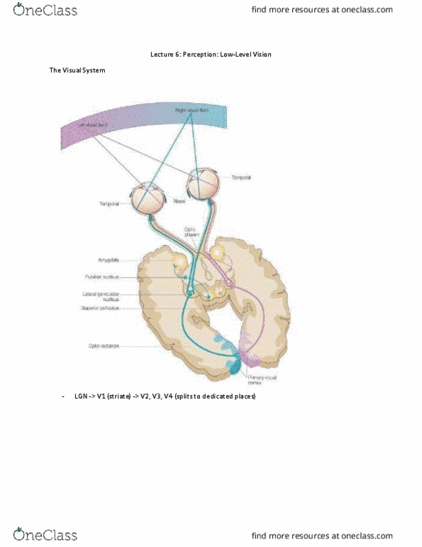
Lgn v1 (striate) v2, v3, v4 (splits to dedicated places) Dorsal pathway where magnocellular spatial relations, movement, etc. Organized spatially two regions close together will project to neurons that are close together in the visual cortex. Watching flashing bullseye pattern injected with radioactive glucose to show neural activity (uptake from active neuron) so it could be exposed to radioactively sensitive film to act as a marker. Right visual field left hemisphere of striate. Areas are spatially relevant (in the same kinda pattern/map) Cortical magnification info processed by the fovea in the center, there is more processing of that information (f). information in your direct line of focus is processed the most. Inverted superior (s) visual field projects to inferior (i) cortex and vice versa, we know the image is switched in the back of the eye. When you present stimuli in different spatial locations, there is corresponding activity in the visual cortex and beyond.