BIOM 3200 Lecture Notes - Lecture 11: Distal Convoluted Tubule, Proximal Tubule, Ultrafiltration (Renal)

1 May 2018
School
Department
Course
Professor

BIOM3200 – Renal System
Pages: 581-615
Renal Physiology*
The kidneys regulate the volume and composition of the body fluids, via a combination of functions:
• They filter blood plasma, separate wastes from the useful chemicals, and eliminate the waste
• They regulate blood volume and pressure by eliminating or conversing water as necessary
• They regulate the osmolarity of the body fluids by controlling relative amounts of water and
solutes eliminated
The kidneys do this by:
• Secreting the enzyme renin, which activates hormonal mechanisms that control blood pressure
and electrolyte balance
• Secreting the hormone erythropoietin which controls the red blood cell count and oxygen-
carrying capacity of the blood
• Functioning with the lungs to regulate the PCO2 and acid-base balance of body fluids
• Contributing to calcium homeostasis through their role in synthesizing calcitrol (vitamin D)
• Detoxifying free radicals and drugs
The kidneys are essential to life
• Loss of kidney function leads to uremia (syndrome of diarrhea, vomiting, dyspnea and cardiac
arrhythmia caused by the toxic effects of nitrogenous wastes)
• Convulsions, coma and death can follow within in a few days
• Unless a kidney transplant is available, renal failure requires hemodialysis to remove nitrogenous
wastes from the blood
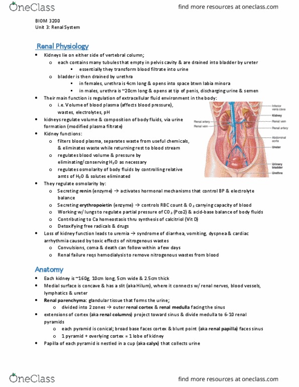
Anatomy*
• Each human kidney weighs ~160g and measures ~10cm long, 5cm wide and 2.5cm thick
o The medial surface is concave and has a slit (the hilum) where it connects with the renal
nerves, blood vessels, lymphatics and ureter
• The renal parenchyma (glandular tissue that forms the urine) is divided into two zone:
o Outer renal cortex (1 cm thick)
o Inner renal medulla facing the sinus
• Extensions of the cortex (=renal columns) project toward the sinus and divide the medulla into 6-
10 renal pyramids
o Each pyramid is conical, with a broad base facing the cortex and a blunt point called the
renal papilla facing the sinus
o One pyramid and the overlying cortex constitute one lobe of the kidney
o The papilla of each renal pyramid is nestled in a cup = minor calyx (which collects its
urine)
o Two or three minor calyces converge to form a major calyx, and two/three major
calcyces converge in the sinus to form the funnel-like renal pelvis
o The ureter is a tubular continuation of the renal pelvis that drains the urine down to the
urinary bladder
• Circulation:
o Although the kidneys account for only 0.4% of the body weight, they receive about 21%
of the cardiac output very important to homeostasis
find more resources at oneclass.com
find more resources at oneclass.com

▪ Each kidney is supplied by a renal artery arising from the aorta
▪ Just before/after entering the hilum, the renal artery divides into a few segmental
arteries, and each of these gives rise to a few interlobar arteries
▪ An interlobar artery penetrates each renal column and travels between the
pyramids toward the corticomedullary junction, the boundary between the cortex
and medulla
▪ Along the way, it branches again to form arcuate arteries, which make a sharp
90 bend and travel along the base of the pyramid
▪ Each arcuate artery gives rise to several inter-lobular arteries which pass upward
into the cortex
o As an interlobular artery ascends through the cortex, a series of afferent arterioles branch
out at right angles
▪ Each afferent arteriole supplies one nephron
▪ It leads to a spheroidal mass to capillaries=glomerulus
▪ The glomerulus is drained by an efferent arteriole
o The afferent and efferent arterioles penetrate one side of the glomerular capsule together
▪ Just outside the capsule, they contact the first part of the distal convoluted tubule
and with it, form a juxtaglomerular apparatus which is critical for the
autoregulation of the kidney
▪ The efferent arteriole usually leads to a plexus of peritubular capillaries, named
for the fact that they form a network around the renal tubules
▪ These capillaries pick up the water and solutes reabsorbed by the renal tubules
▪ From the peritubular capillaries, blood flows into interlobular veins, arcuate
veins, interlobar veins and the renal vein (in that order)
▪ These veins travel parallel to the arteries of the same names
▪ The renal vein leaves the hilum and drains into the inferior vena cava
o The renal medulla receives only 1-2% of total renal flow, supplied by a network of
vessels =vasa recta
▪ These arise from the nephrons in the deep cortex, closest to the medulla
(juxtamedullary nephrons)
▪ Here, the efferent arterioles descend immediately into the medulla and give rise
to the vasa recta instead of peritubular capillaries
▪ The capillaries of the vasa recta lead into venules that ascend and empty into the
arcuate and interlobular veins
• Figure 17.4: Major blood vessels of the kidney
The Nephron*
• Nephron – functional excretory unit of the kidney
o Each human kidney contains ~1.2 million nephrons
• A nephron consists of two principle parts:
o Renal corpuscle – where blood plasma is filtered
▪ Consists of the glomerulus and a two-layered glomerular (Bowman’s) capsule
that encloses it
• The parietal (outer) layer of the capsule is a simple squamous epithelium
and the visceral layer consists of elaborate cells (=podocytes) wrapped
around the capillaries of the glomerulus
• The fluid that filters from the glomerular capillaries (glomerular filtrate)
collects in the capsular space between the parietal and visceral layers and
then flows into the renal tubule on one side of the capsule
find more resources at oneclass.com
find more resources at oneclass.com

▪ The afferent arteriole enters the capsule, bringing blood to the glomerulus, and
the efferent arteriole leaves the capsule and caries it away
• The afferent arteriole is slightly larger than the efferent
• Therefore, the glomerulus has a large inlet and small inlet, which allows
it to autoregulate its own blood pressure
o Long renal tubule – processes this filtrate into urine
▪ This duct leads away from the glomerular capsule and ends at the tip of a
medullary pyramid
• It is ~3cm long and divided into four major regions:
o Proximal convoluted tubule
o Nephron loop (loop of Henle)
o Distal convoluted tubule
o Collecting duct
• First 3 are parts of an individual nephron, the collecting duct receives
fluid from many nephrons
▪ The proximal convoluted tubule (PCT) arises from the glomerular capsule
• Longest and most coiled region
• It has a simple cuboidal epithelium with prominent microvilli
• After coiling extensively near the renal corpuscle, it straightens out and
forms a long U-shaped nephron loop
o Descending limb passes the cortex into the medulla
o Ascending limb returns to the medulla
▪ The nephron loop is divided into thick and thin segments
• Thick – simple cuboidal epithelium; form initial part of the descending
limb and part/all of the ascending limb
o Cells here are heavily engaged in active transport, so they have a
very high metabolic activity and are loaded with mitochondria
▪ When the nephron loop returns to the cortex, it coils again and forms the distal
convoluted tubule
• Shorter and loess convoluted than proximal
• Has cuboidal epithelium with smooth-surfaced cells nearly devoid of
microvilli
• The flow of fluid from point where glomerular filtrate is formed where urine leaves the renal
medulla:
o Glomerular capsule
o Proximal convoluted tubule
o Nephron loop
o Distal convoluted tubule
o Collecting duct
Urine Formation*
Urine formation consists of four main processes:
1. Filtration – fluid and solutes are filtered through the glomerulus and enter Bowman’s capsule
2. Tubular reabsorption recovers useful solutes and water
3. Tubular secretion eliminates wastes from blood
4. Water conservation – water is removed from the filtrate, resulting in urine which then passes into
the collecting ducts
Figure 17.21: Secretion is the reverse of absorption
find more resources at oneclass.com
find more resources at oneclass.com