BIOM 3200 Lecture Notes - Lecture 9: Vascular Endothelial Growth Factor, Vascular Smooth Muscle, Theca Interna

1 May 2018
School
Department
Course
Professor

BIOM3200 – Cardiovascular II
Pages: 431-436 (section 13.6), 450-482 (stop at 14.7)
Blood Vessels
• Thick muscle layer of the arteries allows them to transport blood ejected from the heart under
high pressure
• The thinner muscle of veins allows them to distend when an increased amount of blood enters
them, and their one way valves ensure that blood flows back to the heart
• Capillaries facilitate the rapid exchange of materials between the blood and intestinal fluid
• Blood vessels form a tubular network throughout the body that permits blood to flow from the
heart to all the living cells of the body and then back to the heart
• Blood leaving the heart passes through vessels of progressively smaller diameters:
arteries arterioles capillaries
• Capillaries are microscopic vessels that join arterial flow to venous flow
• Blood returning to the heart from the capillaries passes through vessels of progressively larger
diameters: venules veins
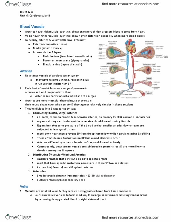
• The walls of the arteries and veins are composed of three coats of “tunics”
o Outermost: tunica externa
▪ Composed of connective tissue
o Middle: tunica media
▪ Composed of smooth muscle
o Inner: tunica interna
▪ Components (inner-outer)
• Inner most simple squamous epithelium (=endothelium) which lines
lamina of all blood vessels
• Basement membrane (layer of glycoproteins) overlying some connective
tissue fibers
• Layer of elastic fibers (=elastin) forming internal elastic lamina
• Although arteries and veins have the same basic structure there are significant differences
o Arteries have more muscle for their diameters than do comparably sized veins
o As a result, arteries appear more rounded in a cross section whereas veins are usually
partially collapsed
o Also, many veins have vales which are absent in arteries
Arteries
• In the aorta and other large arteries there are numerous layers of elastin fibers between smooth
muscle cells of the tunica media
• These large elastic arteries expand when the pressure of the blood rises as a result of ventricle
contraction
o When blood pressure falls, they recoil like a stretched rubber band (when ventricles
relax)
o This elastic recoiling drives the blood during the diastolic phase (longest phase of the
cardiac cycle) when the heart is resting and not providing the driving pressure
• The small arteries and arterioles are less elastic than larger arteries and have a thick layer of
smooth muscle for their diameters
find more resources at oneclass.com
find more resources at oneclass.com

o Therefore, the diameter of the smaller muscular arteries changes only slightly as the
pressure of the blood rises and falls
o Because arterioles and small muscular arteries have narrow lumina, they provide the
greatest resistance to blood flow through the arterial system
• Small muscular arteries that are 100um or less in diameter to form smaller arterioles (20-30um in
diameter)
o In some tissues, blood from the arterioles can enter the venules directly through
atriovenous anastomoses
o In most cases, blood from the arterioles pass into capillaries
o Capillaries are the narrowest of blood vessels (7-10um) – serve as the “business” end of
the circulatory system where gases and nutrients are exchanges between the blood and
tissues
• Resistance to blood flow is increased by vasoconstriction of arterioles (by contraction of smooth
muscle layer) which decreases the blood flow downstream in the capillaries
o Conversely, vasodilation of arterioles (by relaxation of smooth muscle layer) decreases
the resistance and thus increase the flow through the arterioles to the capillaries
• There is evidence of gap junctions between the cells of the arterial wall in both endothelial and
smooth muscle layers
o Vasoconstrictor effect of norepinephrine and the vasodilator effect of acetylcholine may
be propagated for some distance along the arteriole wall by transmissions of
depolarization and hyperpolarization (respectively) through gap junction in vascular
smooth muscle
Capillaries
• The arterial system branches extensively to deliver blood over 40 billion capillaries in the body
o The number of capillary branches is so great that scarcely any cell in the body is 60-80um
away from a blood capillary
o The tiny capillaries provide a total surface area of 1000 square miles for exchanges
between blood and tissue fluid
• The amount of blood flowing through a particular capillary bed depends primarily on the
resistance to blood flow in the small arteries and arterioles that supply blood to capillary bed
o Vasoconstriction in these vessels decreases blood flow, whereas vasodilation increased
blood flow to capillary bed
o The relatively high resistance in the small arteries and arterioles in resting skeletal
muscles, for example, reduces capillary blood flow to only ~5-10% of its maximum
capacity
o In some organs (like intestine), blood flow may be regulated by circular muscle bands
(=precapillary sphincters) at the origin of the capillaries
• Unlike the vessels of the arterial and venous systems, the walls of capillaries are composed of just
one cell layer – a simple squamous epithelium (or endothelium)
o Absence of smooth muscle and connective tissues permits a more rapid exchange of
materials between blood and tissues
• Types of Capillaries:
o Different organs have different types of capillaries, distinguished by significant
differences in structure in terms of endothelial lining:
▪ Continuous capillaries
• Adjacent endothelial cells are closely joined together
• Found in muscles, lungs, adipose tissue and CNS
• The lack of intercellular channels in continuous capillaries within the
CNS contribute to blood-brain barrier
find more resources at oneclass.com
find more resources at oneclass.com

• Continuous capillaries in other organs have narrow intercellular channels
that permit the passage of molecules (other than protein) between blood
and tissue fluid
• Examination of endothelial cells within electron microscope show
presence of pinocytotic vesicles
o Suggests that intracellular transport of materials may occur
across capillary walls
o This type of transport is the only mechanism of capillary
exchange available in CNS
▪ Fenestrated capillaries
• Occur in kidneys, endocrine glands and intestines
• Characterized by wide intercellular pores that are covered by a layer of
mucoprotein (serves as basement membrane over capillary endothelium)
• Mucoprotein layer restricts the passage of certain molecules that might
otherwise pass through pores
▪ Discontinuous capillaries
• Found in bone marrow, liver and spleen
• Distance between endothelial cells is so great that capillaries look like
sinusoids in the organ
• In hypoxic tissues, new capillary networks are stimulated to grow by vascular endothelial growth
factor (VEGF)
o Capillary growth may also be promoted by adenosine (derived from AMP) which
stimulates vasodilation of arterioles and thereby increases blood flow to the hypoxic
tissue
o These changes result in a greater delivery of oxygen-carrying blood to the tissues
Veins
• most of the total blood volume is contained in the venous system
• unlike arteries (which provide resistance to the flow of blood from the heart), veins are able to
expand as they accumulate additional amounts of blood
o vein pressure = 2 mmHg; artery pressure = 100 mmHg
• the low venous pressure is insufficient to return blood to the heart (particularly from lower limbs)
o veins pass between skeletal muscle groups that provide a massaging action as they
contract
o as the veins are squeezed by contracting skeletal muscles, one-way flow of blood to the
heart is ensured by presence of venous valves
• effect of massaging action by skeletal muscle = skeletal muscle pump
o rate of venous return is dependent on the action of pump
o when pumps are less active (individual is still or bedridden), the blood accumulates in the
veins and causes them to bulge
o when pumps are more active (person is active), blood returns to the heart at a faster rate
and less is left in the venous system
• action of the skeletal muscle pump aids the return of venous blood from lower limbs into the
large abdominal veins
o movement of venous blood from abdominal to thoracic veins is aided by breathing
▪ when person inhales, diaphragm (muscular sheet separating thoracic and
abdominal cavities) contracts
▪ contraction of the diaphragm causes it to flatten and descend inferiorly into the
abdomen
find more resources at oneclass.com
find more resources at oneclass.com
Document Summary
Pages: 431-436 (section 13. 6), 450-482 (stop at 14. 7) In some tissues, blood from the arterioles can enter the venules directly through atriovenous anastomoses. In this way, a fall in blood pressure can produce a reflex increase in the heart heart: this baroreceptor reflex is essential for blood pressure regulation. Effects of autonomic nerve activity on the heart: Increased rate of diastolic depolarization increased cardiac rate. In order for this to be true, the strength of ventricular contraction must increase as end-diastolic volume increases. Frank-starling law of the heart: strength of ventricular contraction varies directly with end-diastolic volume, the relationship between edv, contraction strength and stroke volume are intrinsic properties of the heart muscle. Exchange of fluid between capillaries and tissues: distribution of extracellular fluid between plasma and interstitial compartments is in a state of dynamic equilibrium, tissue fluid is not normally a stagnant pond but a continuously circulating medium.