NURS105 Lecture Notes - Lecture 3: Palpebral Fissure, Optic Disc, Lacrimal Gland

5 Mar 2017
School
Department
Course
Professor
Document Summary
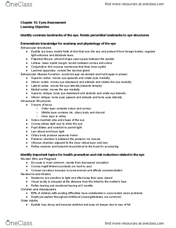
Your objective is to accurately assess visual acuity, extraocular movements, pupilary reaction, and auditory acuity; and to properly perform a fundoscopic exam. Anatomy review eyes: the upper eyelid covers a portion of the iris, but does not overlay the pupil. The opening between the eyelids is called the palpebral fissure: the white sclera may look colored at periphery. Do not mistake this for jaundice, which is a deeper yellow: the conjunctiva is a clear mucous membrane with two easily visible components, bulbar conjunctiva covers most of the anterior eyeball. It adheres loosely to the underlying tissue and meets the cornea at the limbus: the palpebral conjunctiva lines the upper and lower eyelids. The macula surrounds the fovea but has no discernible margins. Note the absence of retinal vessels in much of this area: the retina is the light-sensitive membrane that covers the fundus.