BMS 870 Lecture Notes - Lecture 7: Positron Emission Tomography, Magnetic Resonance Imaging, Choroid Plexus

28 Oct 2017
School
Department
Course
Professor
Document Summary
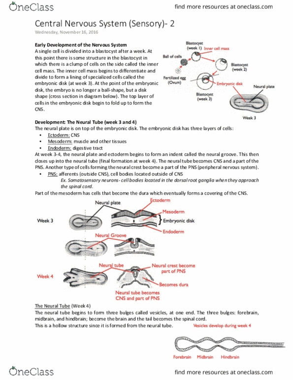
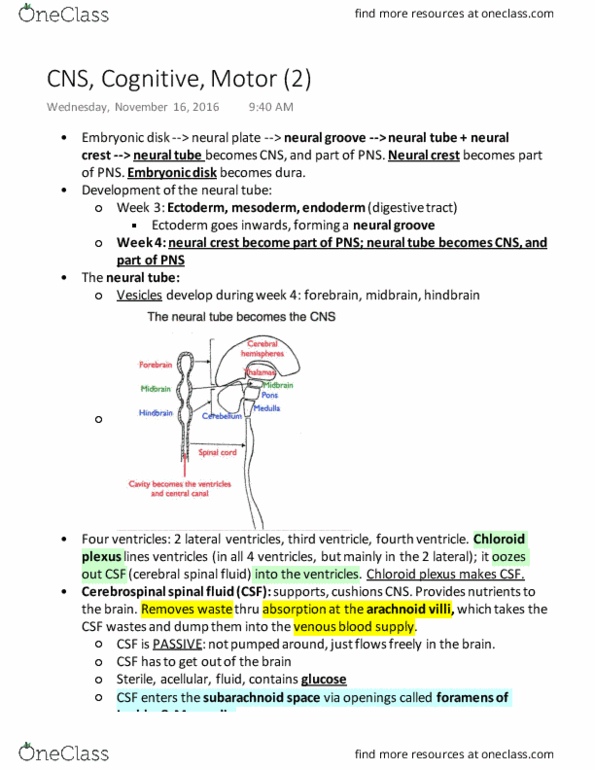
Dorsal root ganglia (drg) - clusters of somas outside of spinal cord that contain somatic sensory axons. Visceral pns - internal organs, blood vessels, glands. Ventricular system = 4 ventricles = 2 lateral ventricles (septa pellucida) + 3rd lateral ventricle + 4th ventricle. Choroid plexus secretes csf which circulates in ventricles, absorbed by arachnoid villi and re-enters blood stream. Circulation of csf: choroid plexus -> fourth ventricle -> central canal of spinal cord -> upward dura mater -> around subarachnoid space -> reabsorbed by arachnoid granulation > into veinous sinus -> leftover in blood. Clarity - transparent brain by replacing lipids w/ water soluble gel. Computed tomography (ct) - x-ray beams creates image. Magnetic resonance imaging (mri) - signals from how h+ perturbations of strong magnetic field. Adv: detailed, no x-ray, image at any angle. Diffusion tensor imaging - detects bundles of axons; image generated by water diffusion (moves easily along path of axon)