MICR 221 Lecture Notes - Lecture 6: Chelation, Ion Trap, Guanidine

12 Nov 2016
School
Department
Course
Professor
Document Summary
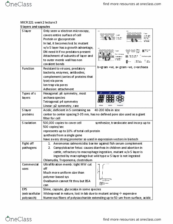
2-d lattice on the surface of a cell envelope like floor tiles. Made of a single protein or glycoprotein. Attached to the cell wall by ionic, hydrophobic or hydrogen bonds (non covalent!) I(cid:374) the la(cid:271), the (cid:858)s(cid:859) la(cid:455)er is ofte(cid:374) (cid:374)ot (cid:374)e(cid:272)essar(cid:455) so the (cid:449)ild t(cid:455)pe (cid:449)ith a(cid:374) s la(cid:455)er are out(cid:374)u(cid:373)(cid:271)ered (cid:271)(cid:455) the (cid:373)uta(cid:374)ts (cid:449)ho do(cid:374)(cid:859)t (cid:373)ake the s la(cid:455)er (cid:271)e(cid:272)ause the(cid:455) ha(cid:448)e a gro(cid:449)th ad(cid:448)a(cid:374)tage! Types of s layer: hexagonal (p6) symmetry, tetragonal (p4) symmetry - square, linear (p2 symmetry) - oblique. Reagents can be used to break the non-covalent bonds to the surface and the s layer will come off. Cell synthesizes, translocates and incorporates its own s layer (moved s layer out to outer surface). But it represents up to 10% of total cell protein. In bacteria the shape of the s layer is determined by the peptidoglycan. (in some archea as well)