ANAT 216 Lecture Notes - Lecture 15: Bronchopulmonary Segment, Pleural Cavity, Serous Membrane

2 Oct 2017
School
Department
Course
Professor
Document Summary
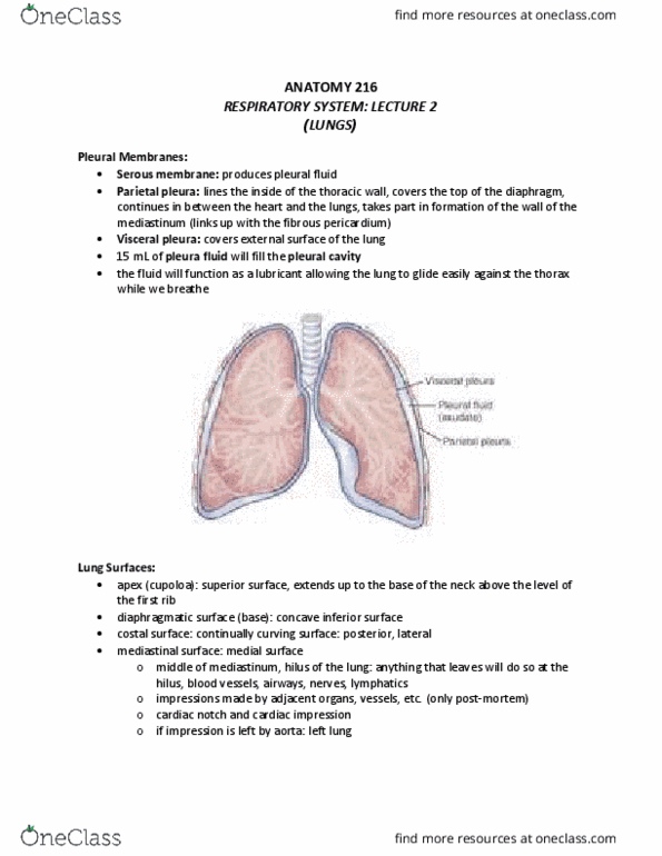
Respiratory system: lecture 2 (reading material: chapter 24: pages 638-647) Cone shaped, each lung is suspended in its own membrane. Serous membrane secretes pleural fluid btwn parietal and visceral membranes to minimize friction btwn lung and wall each time we breathe. Each lung connects by a root (structures coming into or going out of lung) eg. air passageways, parietal (serous membrane, same function as pericardium for heart) visceral covers and stuck tightly to outer surface of lungs cavity, fluid. Covers the top of the diaphragm on each side. Extends btwn heart and lung on each side. Form wall to separate mediastinum from cavity in which lungs are found pulmonary arteries, pulmonary veins, nerves lymphatics. Projects up into base of neck (level of collarbone) Concave surface at base of lung apex (cupoloa) diaphragmatic (base) coastal mediastinal. Continuously cirved surface of anterior, posterior and lateral surfaces. Hilum (indentation in which all structures that form root are located)