ANAT 216 Lecture Notes - Lecture 4: Portal Vein, Common Hepatic Duct, Hepatic Artery Proper

12 Nov 2016
School
Department
Course
Professor
Document Summary
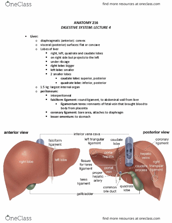
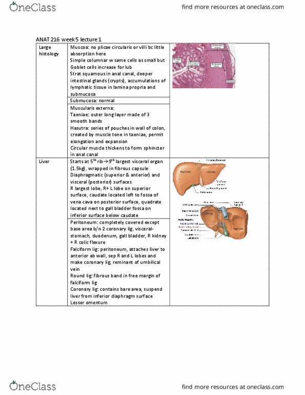
Digestive system: lecture 4 (reading material: chapter 25: pages 682-689) diaphragmatic (anterior) and visceral (posterior) surfaces (faces abdominal) Much of it sit below the right dome of the diaphragm. On the left side sitting under the rib cage. Largest internal organ about 1. 5 kg (in the lab often bigger bc diseased) right, left, quadrate and caudate lobes peritoneum: covers most of liver falciform ligament: round ligament peritoneum between the right and left lobes. Obliterated umbilical vein (carries oxygenated blood to fetus). coronary ligament: - around liver and suspends it. If you cut this there is a part of the liver between the coronary ligament that is touching the diaphragm. Called the care area (liver toughing diaphragm) bare area lesser omentum - porta hepatis (hilus): doorway or gate. Where things enter and leave liver (aka hilus). 3 structures in it portal (hepatic) triad: hepatic artery (proper): (hepatic) portal vein: branch of celiac artery carries oxygenated blood to the liver tissues.