ANAT 215 Lecture Notes - Lecture 5: Tensor Tympani Muscle, Tympanic Duct, Cochlear Duct

28 Apr 2018
School
Department
Course
Professor

ANAT 215 week 2 lecture 2
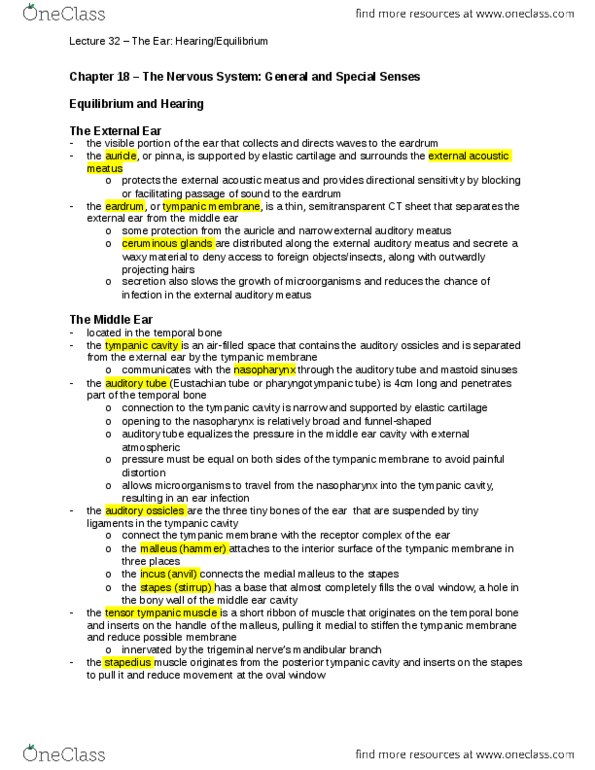
The Ear
Structure
Function
Pic
External
Ear
Auricle/pinna
Cartilage portion,
funnels sound waves
External auditory
canal
Auricle tympanic
memb
Ceruminous gland
Wax gland
Sebaceous
Sweat gland
Tympanic memb
Transmits sound
waves to mech
vibrations
Middle
Ear
Tympanic cavity
Contains ossicles
Pharyngotympanic
tube/ Eustachian
Equalizes pressure,
swallowing opens
tube, pop sound=
tympanic memb
returning to position
Otitis media
Eustachian tube is
shorter + more
vertical= more
infections
Ossicles (malleus
incus stapes oval
window)
Increases sound
intensity to overcome
change in medium
Tensor tympani
muscle (attached to
malleus)
Dampens loud sounds
so they DN damage
hair cells
Stapedius muscle
(attached to stapes)
Dampens loud sounds
so they DN damage
hair cells
Inner
Ear
Labyrinth (bony and
membranous)
Hearing, balance
Endolymph
Fills bony, organ of
corti, cochlear
Perilymph
Fills membranous,
vestibular, tympanic
duct
-
find more resources at oneclass.com
find more resources at oneclass.com