MEDRADSC 2Z03 Lecture Notes - Lecture 5: Nuclear Magnetic Resonance, Magnetic Resonance Imaging, Magnetization

27 Jun 2016
School
Department
Course
Professor
Document Summary
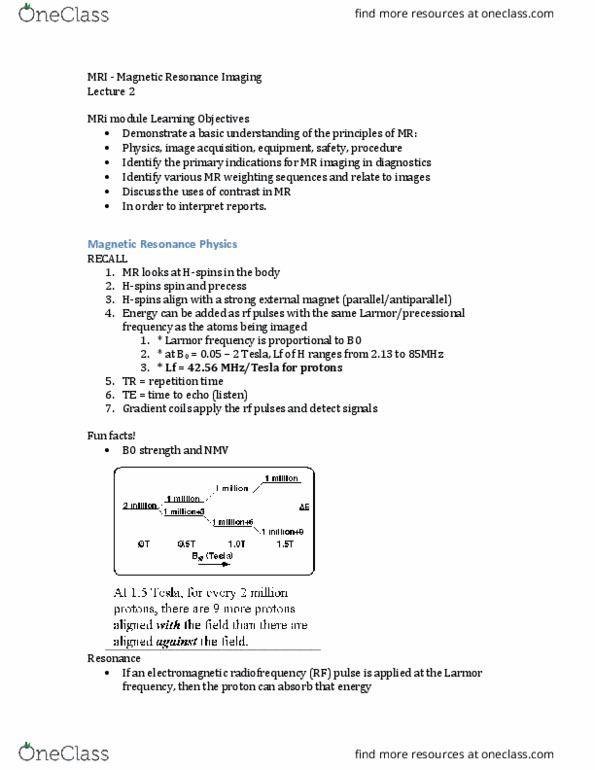
The following slides are a summary of this presentation! Most images are screen captures from the youtube video. Description of the scanning for a patient: Mri = magnetic resonance image/imaging (ie. we don"t say you are getting an mri image taken . Using h-protons that are essentially simple nuclei = nuclear. H spins all have a small magnetic field. Many h-spins in the body (60-75% water, fat, protein) Normally all the little proton-magnets cancel each other out. Add a strong external magnet = magnetization. H-spins align parallel (low e state) or anti-parallel (high e) to the external magnetic field. Creates a nmv net magnetic vector in the longitudinal plane, in the same direction as bo = longitudinal magnetization (cannot be detected) H-spins rotate and precess (rotates like a spin top) Precessional frequency is determined by the larmor frequency and is directly proportional to the external magnetic field strength i. e. 1t magnet precessional (larmor) frequency of h = 42. 58mhz.